目前分類:Cell 細胞 (179)

瀏覽方式: 標題列表 簡短摘要

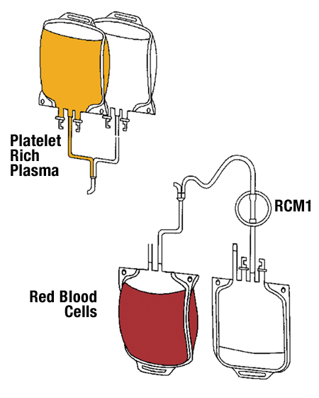
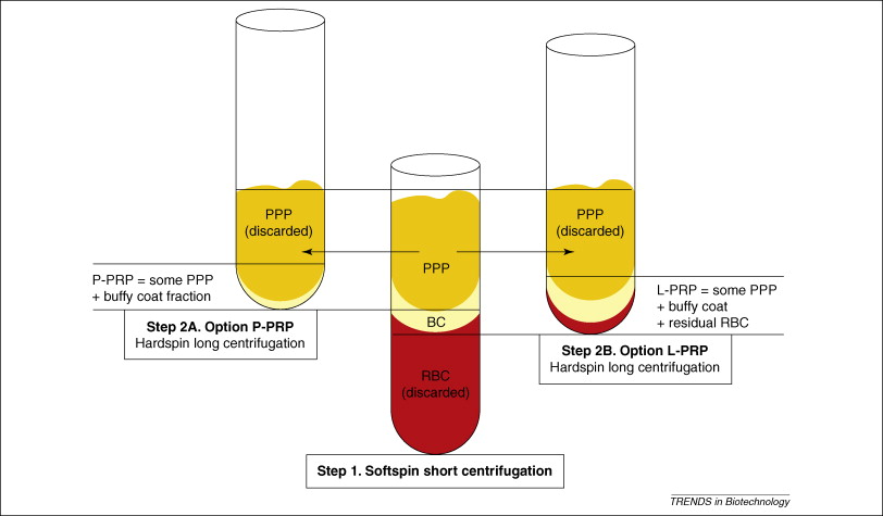
1  1-s2.0-S0167779909000158-gr1  1-s2.0-S0167779909000158-gr3  

L-PRP/L-PRF in Esthetic plastic surgery, Regenerative medicine of the skin and Chronic wounds.

Cieslik-Bielecka A, Choukroun J, Odin G, Dohan Ehrenfest DM.

Curr Pharm Biotechnol. 2012 Jun;13(7):1266-77.


Source

Department and Clinic of Cranio and Maxillofacial Surgery, Medical University of Silesia, ul. Francuska 22-24, 040-096 Katowice, Poland.


Abstract

The use of platelet concentrates for topical use is of particular interest for the promotion of skin wound healing. Fibrin-based surgical adjuvants are indeed widely used in plastic surgery since many years in order to improve scar healing and wound closure. However, the addition of platelets and their associated growth factors opened a new range of possibilities, particularly for the treatment of chronic skin ulcers and other applications of regenerative medicine on the covering tissues. In the 4 families of platelet concentrates available, 2 families were particularly used and tested in this clinical fieldL-PRPLeukocyte- and Platelet-rich Plasmaand L-PRFLeukocyte- and Platelet-Rich Fibrin. These 2 families have in common the presence of significant concentrations of Leukocytes, and these cells are important in the local cleaning and immune regulation of the wound healing process. The main difference between them is the fibrin architecture, and this parameter considerably influences the healing potential and the therapeutical protocol associated to each platelet concentrate technology. In this article, we describe the historical evolutions of these techniques from the fibrin glues to the current L-PRP and L-PRF, and discuss the important functions of the platelet growth factors, the leukocyte content and the fibrin architecture in order to optimize the numerous potential applications of these products in regenerative medicine of the skin. Many outstanding perspectives are appearing in this field and require further research.

Charlie 發表在 痞客邦 留言(0) 人氣()

Do the Fibrin Architecture and Leukocyte Content Influence the Growth Factor Release of Platelet ConcentratesAn Evidence-based Answer Comparing a Pure Platelet-Rich PlasmaP-PRPGel and a Leukocyte- and Platelet-Rich FibrinL-PRF)(3

3. A SIMPLE DEMONSTRATION WITH LARGE PERSPECTIVES.

The release patterns of the 6 tested molecules demonstrate the very significant differences between 2 families of products, P-PRP and L-PRF, and this is therefore an important step for the definitive validation of the current classification of platelet concentrate technologies. Even if both products are platelet concentrates, their intrinsic structure, biology and molecular kinetics are completely opposite. The growth factor release was much more intense in the L-PRF than in the PRGF, but it is not possible to claim that one technique would be better than the other : both families of products can have a potential positive impact during healing, and the 2 technologies have simply different indications since P-PRP are injectable liquid platelet suspensions, while L-PRF only exists as a solid dense fibrin biomaterial.

However, our results suggest that when a fibrin gel biomaterial is needed, L-PRF clots or membranes should be preferred to the PRGF gel : indeed, the L-PRF releases much more Growth factors and key Adhesion proteins during a longer time period and presents a much stronger Fibrin architecture than the PRGF gel. Moreover, the L-PRF technique is very simple and inexpensive, while the PRGF production and clotting technique requires many handling steps and is very time-consuming in order to produce a usable fibrin membrane (clotting during 1 hour at 37deg. Celsius). This conclusion contradicts the published opinion [71] of the PRGF team of the BTI company who claimed that the PRGF technology could be used in many different forms: in the gel form, PRGF is probably not the most adequate platelet concentrate to use, particularly in some applications that require a strong fibrin matrix and simple production procedures of many membranes, such as in oral and maxillofacial surgery.

Logically, this conclusion is true for all P-PRP gels, and not only the PRGF, even if there may be substantial differences in the platelet content between the various P-PRP techniques.

This demonstration of the impact of fibrin architecture and leukocyte content on the biological signature of these products is a first step that opens many perspectives. First, since the various families of product seem to use different intrinsic biological mechanisms, it could be very interesting to investigate properly the effects of these different mechanisms on the proliferation and differentiation of various cell lineages in vitro: the current literature on this topic is often confusing and contradictory because of the lack of proper characterization of the products and associated methodological bias [15]. The following step would be to compare the clinical impact of these 2 families of products. These kinds of comparative studies could increase our knowledge about these products, but remain difficult tasks. Second, our demonstration militates for the refoundation of the current confusing literature about platelet concentrates, by following the simple principles of the classification system [3]. Products should be completely characterized before testing. Fibrin architecture and leukocyte content can not more be neglected in this field.

The perspectives of research are however not only related to platelet concentrates for surgical use. Indeed, blood is a low-cohesion circulating tissue, and it truly gets a full solid tissue cohesion only when it assembles itself in a dense fibrin network during the coagulation process: investigating the mechanisms of L-PRF and P-PRP is therefore not only a way to understand the platelet concentrates technologies, but also a way to understand the blood biology.

Several in vitro articles already demonstrated that the dense Fibrin architecture of a L-PRF membrane allowed a Long slow release of many molecules [14, 68, 72, 73], while the membrane itself was still almost intact after 7 days in the culture medium, even in contact with various cell types in culture [15, 69, 70]: the L-PRF membrane behaves like a true fibrin tissue. L-PRF is often considered as an optimized blood clot [49], and it is indeed a very good illustration of the solid form of the circulating tissue. This slow release was not expected for the products from the PRP families: indeed, these products are brutally activated using bovine thrombin, calcium chloride or other clotting agents, and it was not expected that the platelet growth factors could be trapped intrinsically in this artificial light fibrin network [14, 24, 26, 72]. Our data therefore confirmed the current knowledge about the fibrin clotting [18, 21], and that platelet concentrates may be very interesting models for a better understanding of coagulation and healing mechanisms.

As a conclusion, the key issue in Platelet concentrate technologies is Not the Quantity of platelets, but How Platelet, Leukocytes, Fibrin and Growth factors are interlinked in the final product. A strict quantitative approach does not allow to define the biological signature and mechanisms of a family of platelet concentrates. The approach must be qualitative. PRPs and PRFs are not pharmaceutical preparations with a simple and clear composition, they are living tissues which properties are dependent on the combination of cells, factors and matrix within the final preparation. This demonstration highlights the different mechanisms between 2 families of platelet concentrates, and the significance of the current classification system in order to clarify the literature on the topic and to define adequate research strategies.


DISCLOSURE OF INTEREST

The authors declare no competing financial interests.


ACKNOWLEDGEMENTS

This work was supported by the LoB5 Foundation for Research, Paris, France.


AUTHORS

David M. Dohan Ehrenfest,*, Tomasz Bielecki, Allan Mishra, Piero Borzini, Francesco Inchingolo, Gilberto Sammartino, Lars Rasmusson and Peter A. Everts

Charlie 發表在 痞客邦 留言(0) 人氣()

Do the Fibrin Architecture and Leukocyte Content Influence the Growth Factor Release of Platelet ConcentratesAn Evidence-based Answer Comparing a Pure Platelet-Rich PlasmaP-PRPGel and a Leukocyte- and Platelet-Rich FibrinL-PRF)(2

1. FOUR DIFFERENT FAMILIES OF PLATELET CONCENTRATES, FOUR DIFFERENT BIOLOGICAL MECHANISMS

Since the first articles about platelet concentrate technologies that launched the craze for local application of growth factors [1, 2], many authors tried to compare the characteristics of the various PRP (Platelet-Rich Plasma) techniques available. Indeed, more than 10 different protocols were marketed, and even more in-house protocols were proposed, with various centrifugation and separation procedures, anticoagulant or activators [3].

Many authors tried first to assess the platelet collection efficiency of the various available techniques and the growth factor content of the various products, but they failed to prove a logical correlation between these 2 parameters [4-10]. Three key platelet growth factors were particularly investigated: Transforming Growth Factors 1 (TGF 1), Plate- let-Derived Growth Factors AB (PDGF-AB) and Vascular Endothelial Growth Factors (VEGF). Unfortunately, the literature on growth factor release in PRPs is not very relevant, since most growth factor quantifications were performed on the platelet suspensions before activation, and were therefore meaningless : the true growth factor content could only be assessed after full activation of the product. It was also suspected that many reported data were not using the right scales [11], reporting nanograms/mL (and sometimes micro- grams/mL)[5] of TGF 1 and PDGF-AB, when the normal concentrations for unactivated products should obviously be in picograms/mL.

But the main problem was in fact much deeper : a lack of pertinent terminology and classification led to severe confusions between the many available products and to inadequate methodologies of analysis [12-16]. Most tested products were not fully characterized, and important data such as the leukocyte content [17] and fibrin architecture [18-21] were not assessed [22, 23]. The formation of the fibrin matrix gel during platelet concentrate activation [24] and the leukocyte concentration and formula are obviously key parameters of the growth factor release [11, 25, 26], and also present a strong biological impact in the healing equation [17, 18, 27-29], particularly against infections [30-32]. Without the control of these parameters, the contradictory literature about the effects of the platelet concentrates was mostly focused on the platelet count, and was therefore often biased.

Recently, a full classification of platelet concentrate technologies was designed [3], and allowed to classify the main available techniques in 4 families, depending on their leukocyte content and fibrin architecture :

Pure Platelet-Rich Plasma (P-PRP) and Leukocyte- and Platelet-Rich Plasma (L-PRP) are Liquid platelet suspensions, respectively without and with leukocytes. They can be used as injectable suspension, particularly in sports medicine [33, 34]. After activation (with thrombin, calcium chloride, batroxobin or others agents), these preparations become respectively P-PRP and L-PRP fibrin gels, with a brutal and incomplete fibrinogen polymerization and a Light final fibrin architecture.

Pure Platelet-Rich Fibrin (P-PRF) and Leukocyte- and Platelet-Rich Fibrin (L-PRF) are Solid fibrin biomaterials, respectively without and with leukocytes. In these techniques, the platelet activation is part of the production process: it can be natural (L-PRF) or artificial (P-PRF) but always occurs during the centrifugation, and leads to a Strong final fibrin architecture.

The concept of this classification is to define and regroup the products through their main features and associated biological mechanisms. This is only a first step, since platelet concentrations, leukocyte concentration and formula can also vary within a family of products. This classification is very attractive and logical from a strict theoretical standpoint, but it is now necessary to highlight and clarify the different biological properties and mechanisms associated to each family.


2. EVALUATION OF THE IMPACT OF THE FIBRIN MATRIX AND THE LEUKOCYTE CONTENT ON THE GROWTH FACTOR RELEASE.

2.1. Definition of Anitua’s PRGF (P-PRP) and Choukroun’s PRF (L-PRF)

In order to understand the different biological mechanisms of the 4 families of platelet concentrates, the first step is to compare the growth factor release of well-characterized products from 2 different families. Two products seemed the adequate examples.

The first tested product is a P-PRP called PRGF, aka Plasma [35] or Preparation [36] Rich in Growth Factors. This manual procedure was invented by Anitua in 1999 [35] and is currently marketed by BTI, aka BioTechnology Institute (Vitoria, Spain), a dental implant company directed by Dr Anitua [13]. After a Soft centrifugation of blood with Anticoagulant, Four layers appear in the tube : the superficial plasma suspension was often called PPGF (Plasma Poor in Growth Factors), the intermediate plasma suspension is called PRGF (Plasma Rich in Growth Factors), the whitish layer below the PRGF is called the Buffy coat, and the Red blood cells are finally gathered at the bottom of the tube. The PPGF and the PRGF are collected by pipetting the plasma solution above the red blood cell base. The specificity of this technique is that the Buffy coat layer (between the red blood cells base and the acellular plasma) is Not harvested in order to avoid the collection of Leukocytes [37]. Since the Buffy coat layer contains most Leukocytes and also many platelets, the final PRGF is a Platelet-rich plasma suspension with almost no Leukocyte and a lower platelet concentration than other PRPs [38]. The PRGF suspension can be used like an Injectable pharmaceutical solution, particularly in sports medicine [39] or for the Coating of implant surface [40, 41](even if this last application is very debatable)[42], or can be activated with Calcium chloride in order to prepare a Fibrin gel. This fibrin gel is quite fragile and unstable, and can be used as a fibrin covering layer, like a fibrin glue [37, 43].

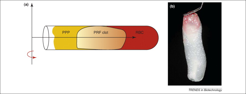
The second tested product is a L-PRF, called Choukroun’s PRF. This open-access technique was invented in 2000 by Choukroun [44], and adequate kits and centrifuge were marketed by Process (Nice, France). The general concept of this technique is very different from the numerous other protocols and was often considered as a second generation platelet concentrate technology [45-48]. Blood is taken Without anticoagulant and immediately Softly centrifuged. Blood activation occurs during the centrifugation and allows the formation of a dense Fibrin-platelet clot in the middle of the tube, between the Red blood cell base at the bottom and the Acellular plasma at the top of the tube. The L-PRF clot contains Almost all the Platelets and more than 50% of the Leukocytes from the initial blood harvest, and presents a strong Fibrin architecture and a specific Three-dimensional distribution of the Platelets and Leukocytes [16, 49]. This product therefore only exists in an activated form and can not be injected like a suspension. Because of its strong fibrin architecture, this solid biomaterial is particularly useful in oral and maxillofacial surgery [50-53], periodontology [54, 55], implant dentistry [56-62] and ENT (Ear Nose Throat) surgery [63-65], and many other applications may be investigated in the future.

PRGF gel and Choukroun’s PRF are therefore the 2 extreme opposites of the platelet concentrate technologies, respectively a P-PRP gel and a L-PRF. The 2 materials can easily be prepared in similar solid forms and volumes, and the comparison of their growth factor release is of great interest in order to understand the impact of the leukocyte content and the fibrin architecture on the intrinsic biological mechanisms of these living biomaterials.


2.2. Comparison of the Protein Release Patterns

In a recent study, it was proven that a L-PRF membrane slowly releases significant amounts of some growth factors (TGF 1, PDGF-AB, VEGF) and thrombospondin-1 (TSP-1) during at least 7 days [11]. The method was simple: the L- PRF membrane was placed in a 10 mL tube with 4 mL of sterile Dulbecco’s modified eagle’s medium (DMEM). Then, at each experimental time, the membrane was transferred in a new tube with 4 mL sterile DMEM, and the previous 4 mL were stored at -80 deg. Celsius before ELISA quantification. The membrane transfer was done at seven experimental times: 20 min, 1h, 4h, 24h (day 1), 72h (day 3), 120h (day 5) and 168h (day 7). The initial growth factor content of a L-PRF membrane after forcible extraction was also quantified. The comparison between the 7-day slow released quantities and the initial growth factor content highlighted interesting data: the PDGF-AB initial quantities were quite similar to the final released amounts after 7 days. But the released amounts of TGF 1 and VEGF after 7 days were more than 6 times higher than the initial content. It was thus hypothesized that the Leukocytes trapped within the L-PRF membranes were producing these growth factors large surplus.

Following exactly the same protocol and using the same four volunteer blood donors than in the first article (two males and two females, non-smokers, aged between 50 and 55 years old), we decided to evaluate the release of the 3 same Growth factors (TGF 1, PDGF-AB, VEGF) and 3 key matrix proteins (Thrombospondin-1, Fibronectin and Vitronectin) from a L-PRF membrane and from a PRGF gel membrane. Fibronectin and Vitronectin are 2 key Cell adhesion and migration proteins. The fibronectin is present in great quantities in a blood circulating form and in the platelets [66], and is also a key component of the architecture of the fibrin clot [67]. Therefore, the Fibronectin released from a platelet concentrate gel is first the soluble free fibronectin, and not the fibronectin blocked in the in-depth architecture of the fibrin matrix.

Choukroun’s PRF (Process, Nice, France) and Anitua’s quantifications of growth factors and matrix proteins were performed in triplicate with ELISA kits (Quantikine, R&D Systems, Minneapolis, MN, USA, for TGF 1, PDGF-AB, VEGF and Thrombospondin-1 ; EIA kit, GenWay, San Di- ego, CA, USA, for fibronectin and vitronectin).

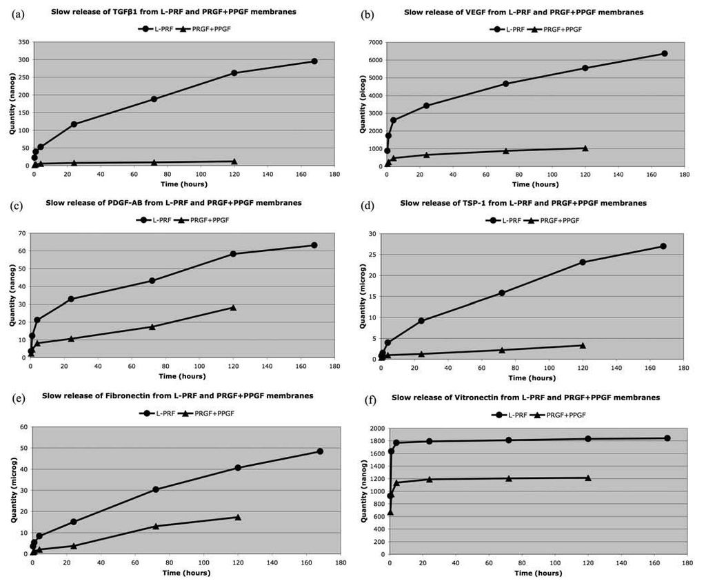
The results are reported in the Table 1 and in the Fig. (2), and demonstrate the impact of the cell content and the fibrin architecture in the biology of these platelet concentrates.

First, after 20 minutes the release of Growth factors and Matrix proteins from the L-PRF is always very significantly (p<0.0001) superior to the release of the PPGF and PRGF membranes together, during the 7 days of experiment.

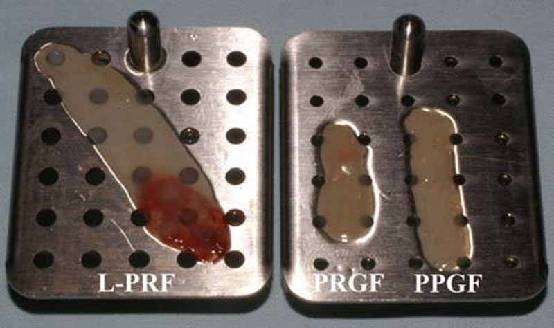
Second, at the 5th day of the experiment, the PPGF and PRGF membranes were completely dissolved in the culture medium. The 2 membranes looked already damaged on the PRGF (BTI, Vitoria, Spain) were produced with their official third day, and disappeared between the 3rd and the 5th days of kits and following the classical protocols previously described in the literature. For the Choukroun’s PRF, blood was harvested in 9mL glass-coated plastic tubes, and the L- PRF clot was directly collected at the end of the centrifugation. For Anitua’s PRGF, blood was collected in two 4.5mL Citrated tubes, and after centrifugation, the 2 PPGF were gathered in a tube and the 2 PRGF gathered in another tube for Separate clotting by adding Calcium chloride and waiting a dense Polymerization for 1 h at 37deg. Celsius. All the membranes were finally prepared and standardized using the PRF Box (Process, Nice, France), an user-friendly device that allows to compress the PRF and PRGF/PPGF clots into membranes in a sterile and protected environment [68]. Finally, with the same 9mL blood volume, we produced one big L-PRF membrane and 2 small P-PRP gel membranes (one PRGF and one PPGF). The PRGF and PPGF membranes together presented almost the same volume than a L-PRF membrane alone

Fig. (1). The comparison of the growth factor releases from the L-PRF membrane and the PPGF/PRGF gel membranes is therefore particularly relevant. Three L-PRF and three PPGF/PRGF membranes were produced for each donor. The experiment. On the contrary, the L-PRF membrane seemed intact after 7 days in culture, as it was reported previously in various in vitro culture studies [69, 70], and the L- PRF could probably remain in the experiment even longer. This observation confirmed that the fibrin architecture is much stronger in the PRF subfamilies than in the PRP gel classes.

Third, the growth factor releases of the PPGF membranes were not significantly different from the releases of the PRGF membranes; this result was associated to very large standard deviations of the measured mean quantities of released proteins from the PRGF and PPGF. This observation was quite surprising, since the PPGF was supposed to present a lower platelet concentration than the PRGF. However, there is a simple explanation : the PRGF technique is a manual procedure that requires several steps of pipetting with eye-balling a sole measuring method. The separation of the PPGF and PRGF layers in the plasma supernatant is very theoretical, since the buffy coat is not collected. Pipetting in a small tube always creates turbulences that unavoidably homogenize partially the PPGF and PRGF layers. Our results therefore demonstrate that the PPGF and PRGF layers present similar biological signatures, and that their theoretical separation is an empirical concept.

Finally the release patterns were very different between the L-PRF and the PPGF/PRGF membranes, and give us considerable information about the effect of the fibrin architecture and the leukocyte content on the mechanisms of the release. The L-PRF membrane slowly released large amounts of TGF 1, PDGF-AB, VEGF, Thrombospondin-1 and Fibronectin during at least 7 days. If the stronger release occurs during the first 24 hours, the membrane released large amounts continuously during the whole experiment. On the contrary, in PRGF/PPGF membranes, TGF 1, PDGF-AB, VEGF and Thrombospondin-1 were mainly released during the first 4 hours and, even if a small release continued up to the final dissolution of the PPGF/PRGF membranes, this slow release was considerably less intense than in a L-PRF membrane (this can be easily observed on the slope of the release curves in the Fig. (2). The situation was even more obvious with the fibronectin, that was released in 2 phases from the PRGF/PPGF membranes : half of the total content was released massively during the first hour, and the remaining fibronectin was released between the 3rd and 5th days when the PPGF/PRGF membranes finally dissolved. The explanation of these different release patterns is quite simple: during the platelet and fibrinogen activation of PPGF/PRGF, growth factors and some other proteins are not enmeshed in the fibrin network, because the fibrin polymerization is incomplete. These molecules are therefore released massively during the first hours after preparation. Moreover, these products do not contain leukocytes and can therefore not sustain the production of new growth factors after the initial release. On the contrary, the strong fibrin architecture of the L-PRF allows an intense slow release during the whole experiment, and the release is even supported by the production of new growth factors (particularly TGF 1) by the leukocytes living in the L-PRF membrane.

There is only one exception to these release patterns : in L-PRF and PPGF/PRGF membranes, the Vitronectin was massively released during the first 4 hours, and the release was then almost nil during the next 7 days. This observation demonstrates that some molecules can not be trapped in the fibrin matrix, whatever the method of polymerization, and are released almost immediately after production. It also means that if we need Vitronectin on a surgical site, the membranes should be used quite Quickly after preparation (even if the conservation in a PRF Box may probably delayed the vitronectin release) or we should also use the clot exudate collected in the PRF Box [68].

Charlie 發表在 痞客邦 留言(0) 人氣()

 1  2  

Do the Fibrin Architecture and Leukocyte Content Influence the Growth Factor Release of Platelet ConcentratesAn Evidence-based Answer Comparing a Pure Platelet-Rich PlasmaP-PRPGel and a Leukocyte- and Platelet-Rich FibrinL-PRF)(1

http://www.francescoinchingolo.it/default.asp?modulo=news&action=read&id=158


Platelet concentrates for surgical use are tools of regenerative medicine designed for the local release of platelet growth factors into a surgical or wounded site, in order to stimulate tissue healing or regeneration. Leukocyte content and Fibrin architecture are 2 key characteristics of all platelet concentrates and allow to classify these technologies in 4 families, but very little is known about the impact of these 2 parameters on the intrinsic biology of these products. In this demonstration, we highlight some outstanding differences in the growth factor and matrix protein release between 2 families of platelet concentrate : Pure Platelet-Rich Plasma (P-PRP, here the Anitua’s PRGF - Preparation Rich in Growth Factors - technique) and Leukocyte- and Platelet-Rich Fibrin (L-PRF, here the Choukroun’s method). These 2 families are the extreme opposites in terms of fibrin architecture and leukocyte content. The Slow release of 3 key Growth factors (Transforming Growth Factor 1 (TGF 1), Platelet-Derived Growth Factor AB (PDGF-AB) and Vascular Endothelial Growth Factor (VEGF) and Matrix proteins (Fibronectin, Vitronectin and Thrombospondin-1) from the L-PRF and P-PRP gel membranes in culture medium is described and discussed. During 7 days, the L-PRF membranes Slowly release significantly Larger amounts of all these molecules than the P-PRP gel membranes, and the 2 products display different release patterns. In both platelet concentrates, Vitronectin is the sole molecule to be released almost completely after only 4 hours, suggesting that this molecule is not trapped in the fibrin matrix and not produced by the leukocytes. Moreover the P-PRP gel membranes completely dissolve in the culture medium after less than 5 days only, while the L-PRF membranes are still intact after 7 days. This simple demonstration shows that the polymerization and final architecture of the fibrin matrix considerably influence the strength and the growth factor trapping/release potential of the membrane. It also suggests that the Leukocyte populations have a strong influence on the release of some growth factors, particularly TGF-1. Finally, the various platelet concentrates present very different biological characteristics, and an accurate definition and characterization of the different families of product is a key issue for a better understanding and comparison of the reported clinical effects of these surgical adjuvants.


Fig. (1). L-PRF and PRGF/PPGF membranes just after preparation in the PRF Box. The membranes are ready for insertion in the test tubes. The L-PRF membrane was produced with the same quantity of blood (9mL) than the PRGF and PPGF membranes together, and the final size and volume of the 2 platelet preparations (L-PRF and PRGF+PPGF) was comparable. However, the PRGF or PPGF membrane alone is obviously smaller and more fragile than the L-PRF membrane.

Fig. (2). Slow release of TGF 1 (a), VEGF (b), PDGF-AB (c), Thrombospondin-1 (d), Fibronectin (e) and Vitronectin (f) from a L-PRF membrane and PRGF+PPGF membranes during 7 days. Values are expressed as the cumulative mean quantity of molecules at 20 minutes, 1 hour, 4 hours, 24 hours, 72 hours (3 days), 120 hours (5 days) and 168 hours (7 days).

Charlie 發表在 痞客邦 留言(0) 人氣()

jcad_4_12_32-g005  jcad_4_12_32-g006  jcad_4_12_32-g008  

Avoiding Malar Edema During Midface/Cheek Augmentation with Dermal Fillers3

The final injection (not shown) is at the medial portion of the zygomatic arch. At each site, the needle is walked along the periosteum, depositing small amounts of filler without withdrawing the needle to limit the number of puncture sites and their resultant ecchymosis and edema. HA-JU is injected with a 30-gauge, ½-inch needle. CaHA is injected with a 28-gauge, ¾-inch needle.

The material is molded to smoothness gently so as not to predispose the patient to ecchymoses. The purpose of Molding is to smooth and manipulate the filler into the area of volume deficiency. Attention is directed when molding not to overly flatten or disperse the filler, necessitating higher volume of filler as a result of loss of correction. Overzealous massage can result in filler moving more superficially through needle tracts, thus increasing the propensity for visible material and malar edema. The ultimate objective is a Smooth blending between the lower eyelid, nasolabial fold, and the cheek. All bleeding points are treated with immediate and sustained direct pressure. Postinjection ice packs and Head elevation are employed.

Midfacial volume restoration using fillers is performed Medially to Laterally, since Volumes should be most Limited Medially beneath the malar septum. Volumes need only be restricted by aesthetic goals when treating the malar eminence, lateral orbital rim, zygomatic arch, and submalar hollow. Facial volume restoration is performed correcting the Midface prior to the Lower face. Expansion of the midfacial soft tissue envelope will result in an Effacement of the nasolabial folds and a reduction of the filler volume required for their correction.

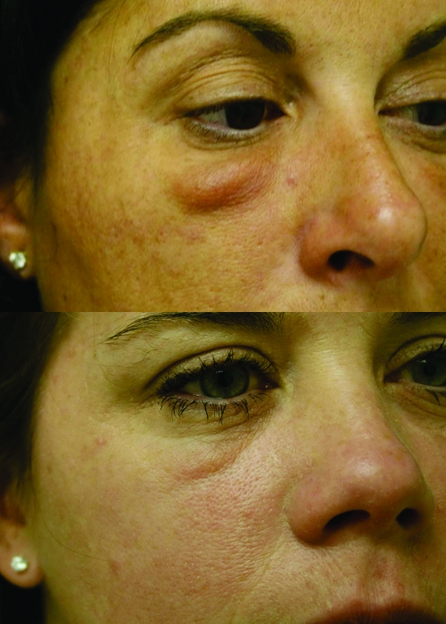
Figures 5 and and66 show three patients with malar edema who had been treated for volume enhancement using HA or CaHA, both injected through a transcutaneous approach using Fanning and Threading technique in the Suborbicularis plane. In Figure 5, a 39-year-old female received HA into her tear troughs and infraorbital rims. She was treated with 20 units of Vitrase (ISTA Pharmaceuticals, Inc.) to ameliorate her malar edema. In Figure 6, a 44-year-old woman received a total of 1.3mL of CaHA in her midface (0.65mL per side), with malar edema evident five weeks post-treatment (A), and a 40-year-old woman received a total of 2.6mL of CaHA (1.3mL per side), with malar edema evident at three weeks post-treatment (B).


Figure 5A and 5B

A 39-year-old woman received hydroxylapatite in her tear trough and infraorbital rim. Malar edema could be observed three weeks post injection (A). The patient was treated with 20 units of Vitrase, which led to resolution (B).


Figure 6A and 6B

A 44-year-old woman received a total of 1.3mL of calcium hydroxylapatite in her midface (0.65mL per side), with malar edema evident five weeks post-treatment (A). A 40-year-old woman received a total of 2.6 mL of calcium hydroxylapatite (1.3mL per side), ...


The author has performed more than 350 midfacial augmentations using this technique without any occurrence of malar edema or other significant adverse events, such as severe bruising, contour irregularities, visible material, or infraorbital nerve injury. The majority of patients were treated with a combination of CaHA and HA-JU. Average volume was one Half a syringe of each material Per side, i.e., 0.65mL of CaHA per side (in 1.3mL syringe) and 0.4mL of HA-JU per side. The HA-JU was injected in the area Beneath the Malar septum and the CaHA for Enhancement of the Malar eminence Lateral to the Lateral canthus. This approach was selected because of the ability to dissolve hyaluronic acid using hyaluronidase, if malar edema or other adverse event should occur. In addition, HA-JU has a significantly lower G' than CaHA, making it less likely to compress the lymphatics of the area bounded by the malar septum. The higher G', or Lifting force exerted by CaHA, the more successfully elevated the thicker cheek tissues of the Malar eminence, Lateral orbital rim, and Zygoma. Figure 7 is representative of

the results obtained in the use of HA in the treatment of tear trough and infraorbital hollow.


Figure 7

Correction of tear trough and infraorbital hollow using calcium hydroxylapatite.


Recently, the author has employed the HA Belotero Basic (Merz), approved in Europe and recently approved in the United States, placed subcutaneously, for correction of tear troughs and infraorbital hollows, without adverse events. No Tyndall effect or visible material was observed. This HA was again combined with CaHA treatment of the malar eminence and inframalar hollow as aesthetically necessary.


Conclusion

Facial volume restoration and contour enhancement using dermal fillers have become a valuable addition to the aesthetic surgeon's armamentarium. These techniques are relatively quick to perform, have little down time, and result in a high rate of patient satisfaction. Adverse events have been reported however, particularly when the area of the lower eyelid are injected. Although malar edema is a severe adverse event, its incidence can be reduced by proper patient selection, proper filler selection, limiting filler volume, and by placing filler material deep into the Malar septum at the immediate Preperiosteal level.


Acknowledgment

The author appreciates the editorial assistance of David J. Howell, PhD, RRT (San Francisco, California) in the development of this manuscript.


Footnotes

DISCLOSURE :

Dr. Funt has received consulting fees for his work with Merz Aesthetics and serves as one of the company's medical advisors. He is also part of the national speakers bureau of Allergan Corporation and receives honoraria for educational activities. Financial support for preparation of this manuscript was provided in part by Merz Aesthetics (San Mateo, California).


REFERENCES

1. Tzikas TL. A 52-month summary of results using calcium hydroxylapatite for facial soft tissue augmentation. Dermatol Surg. 2008;34:S9–315. [PubMed]

2. Graivier MH, Bass LS, Busso M, Jasin ME, Narins RS, Tzikas TL. Calcium hydroxylapatite (Radiesse) for correction of the mid- and lower face: consensus recommendations. Plast Reconstr Surg. 2007;120(Suppl):55S–66S. [PubMed]

3. Silvers SL, Eviatar JA, Echavez MI, Pappas AL. Prospective, open-label, 18-month trial of calcium hydroxylapatite (Radiesse) for facial soft-tissue augmentation in patients with human immunodeficiency virus-associated lipoatrophy: one-year durability. Plast Reconstr Surg. 2006;118(Suppl.):34s–45s. [PubMed]

4. Werschler WP. Treating the aging face: a multidisciplinary approach with calcium hydroxylapatite and other fillers, part 2. Cosmetic Dermatol. 2007;20(12):791–796.

5. Busso M, Karlsberg PL. Cheek augmentation and rejuvenation using injectable calcium hydroxylapatite (Radiesse®) Cosmetic Dermatol. 2006;19:583–588.

6. Pessa JE, Garza JR. The malar septum: the anatomic basis of malar mounds and malar edema. Aesthetic Surg J. 1997;17(1):11–17. [PubMed]

7. Pessa JE, Zadoo VP, Adrian EK, Woodwards R, Garza JR. Anatomy of “black eye”: A newly described fascial system of the lower eyelid. Clin Anat. 1998;11:157–161. [PubMed]

8. Busso M, Voigts R. An investigation of changes in physical properties of injectable calcium hydroxylapatite in a carrier gel when mixed with lidocaine and with lidocaine/ epinephrine. Dermatol Surg. 2008;34:S16–S24. [PubMed]

Charlie 發表在 痞客邦 留言(0) 人氣()

prpFig1  

Platelet-rich plasma preparation for Regenerative medicineoptimization and quantification of Cytokines and Growth factors.

Amable PR, Carias RB, Teixeira MV, da Cruz Pacheco I, Corrêa do Amaral RJ, Granjeiro JM, Borojevic R.

Stem Cell Res Ther. 2013 Jun 7;4(3):67. [Epub ahead of print]


Source

Excellion Biomedical Services, Petrópolis, Rio de Janeiro, Brazil. paola@excellion.com.br.


Abstract

INTRODUCTION :

Platelet-rich plasma (PRP) is nowadays widely applied in different clinical scenarios, such as orthopedics, ophthalmology and healing therapies, as a growth factor pool for improving tissue regeneration. Studies into its clinical efficiency are not conclusive and one of the main reasons for this is that different PRP preparations are used, eliciting different responses that cannot be compared. Platelet quantification and the growth factor content definition must be defined in order to understand molecular mechanisms behind PRP regenerative strength. Standardization of PRP preparations is thus urgently needed.


METHODS :

PRP was prepared by centrifugation varying the relative centrifugal force, temperature, and time. Having quantified platelet recovery and yield, the two-step procedure that rendered the highest output was chosen and further analyzed. Cytokine content was determined in different fractions obtained throughout the whole centrifugation procedure.


RESULTS :

Our method showed reproducibility when applied to different blood donors. We recovered 46.9 to 69.5% of total initial Platelets and the procedure resulted in a 5.4-fold to 7.3-fold increase in Platelet concentration (1.4 × 10⁶ to 1.9 × 10⁶ platelets/μl). Platelets were highly purified, because only <0.3% from the initial red blood cells and leukocytes was present in the final PRP preparation. We also quantified Growth factors, Cytokines and Chemokines secreted by the concentrated platelets after activation with Calcium and Calcium/Thrombin. High concentrations of platelet-derived growth factor, endothelial growth factor and transforming growth factor (TGF) were secreted, together with the Anti-inflammatory and Proinflammatory Cytokines Interleukin (IL)-4, IL-8, IL-13, IL-17, tumor necrosis factor (TNF) and interferon (IFN). No cytokines were secreted before platelet activation. TGF-β3 and IFNγ were not detected in any studied fraction. Clots obtained after platelet coagulation retained a high concentration of several growth factors, including platelet-derived growth factor and TGF.


CONCLUSIONS :

Our study resulted in a consistent PRP preparation method that yielded a cytokine and growth factor pool from different donors with high reproducibility. These findings support the use of PRP in therapies aiming for tissue regeneration, and its content characterization will allow us to understand and improve the clinical outcomes.

Charlie 發表在 痞客邦 留言(0) 人氣()

Platelet-released growth factors enhance the secretion of Hyaluronic acid and induce Hepatocyte growth factor production by Synovial fibroblasts from arthritic patients

E. Anitua1, M. Sánchez2, A. T. Nurden3, M. M. Zalduendo1, M. de la Fuente1, J. Azofra2 and I. Andía1

1Biotechnology Institute, BTI IMASD, 2Unidad de Cirugia Artroscópica ‘Mikel Sanchez’, Vitoria, Spain and 3IFR4/FR21, Pessac, France.

Correspondence to : I. Andia, Biotechnology Institute, c/ Leonardo Da Vinci 14, 01510 Miñano, Alava, Spain. E-mail: isabel.andia@bti-imasd.com

Received May 31, 2007.

Revision received August 2, 2007.


Abstract

Objectives.

Autologous platelet-secreted growth factors (GFs) may have therapeutic effects in osteoarthritis (OA) capsular joints via multiple mechanisms. Our aim was to examine the effect of a platelet-derived preparation rich in growth factors (PRGFs) in OA synovial cell biology.


Methods.

Synovial cells were isolated from 10 osteoarthritic patients and cultured in serum-free media (basal conditions) and exposed to either a platelet-poor preparation or PRGF for 72 h. Cells activated with interleukin-1β (IL-1β) for 48 h were also exposed to PRGF. Changes in several events relevant to joint homeostasis including (i) hyaluronic acid (HA) secretion, (ii) the balance between metalloproteinase-1, -3 and -13 (MMP-1, MMP-3 and MMP-13) and tissue inhibitor-1 (TIMP-1) and (iii) the secretion of transforming growth factor-β1(TGF-β1), vascular endothelial growth factor (VEGF) and hepatocyte growth factor (HGF), were all assessed.


Results.

PRGF significantly enhanced HA secretion compared with platelet-poor preparations, P < 0.05; at the same time release of TIMP-1, MMP-1, MMP-3 and MMP-13 were not affected. An increased HGF production was observed (P < 0.05) but VEGF and TGF-β1 levels remained unchanged. PRGF significantly enhanced the secretion of HA induced by IL-1β activation, P < 0.05, but it did not modify the IL-1β-induced rise in MMP-1, MMP-3 and VEGF. In contrast, PRGF-induced HGF production was abolished by the presence of IL-1β during PRGF treatment, P < 0.05.


Conclusions.

Intra-articular administration of PRGF might be beneficial in restoring HA concentration and switching angiogenesis to a more balanced status but does not halt the effects of IL-1β on synovial cells.


Key words

Platelet-rich plasma Osteoarthritis Synovial cells IL-β Growth factors

© The Author 2007.

Published by Oxford University Press on behalf of the British Society for Rheumatology.

All rights reserved.

For Permissions, please email: journals.permissions@oxfordjournals.org

Charlie 發表在 痞客邦 留言(0) 人氣()

article-2079461-0F49EEC400000578-879_468x286  dunnys-ankle-597x403  kobe-bryant-tweet-ankle  grant_e_centrifuge_576  

Charlie 發表在 痞客邦 留言(0) 人氣()

Blood-centrifugation-scheme  

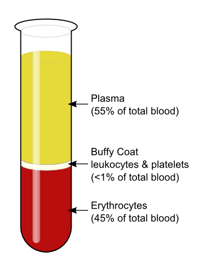
Buffy coatBC):White blood cells and Platelets

From Wikipedia, the free encyclopedia


The buffy coat is the fraction of an anticoagulated blood sample that contains most of the White blood cells and Platelets following density gradient centrifugation of the blood.

After centrifugation, one can distinguish a layer of clear fluid (the plasma), a layer of red fluid containing most of the red blood cells, and a thin layer in between. Making up less than 1% of the total volume of the blood sample, the buffy coat (so-called because it is usually buff in hue), contains most of the White blood cells and Platelets. The buffy coat is used, for example, to extract DNA from the blood of mammals (since mammalian red blood cells are anucleate and do not contain DNA).

The buffy coat is usually Whitish in color, but is sometimes Green if the blood sample contains large amounts of Neutrophils (which are high in green myeloperoxidase). The layer next to buffy coat contains granulocytes and red blood cells.

Quantitative buffy coat (QBC) is a laboratory test to detect infection with malaria or other blood parasites. The blood is taken in a QBC capillary tube which is coated with acridine orange (a fluorescent dye) and centrifuged; the fluorescing parasites can then be observed under ultraviolet light at the interface between red blood cells and buffy coat. This test is more sensitive than the conventional thick smear and in > 90% of cases the species of parasite can also be identified.

In cases of extremely low white blood cell count, it may be difficult to perform a manual differential of the various types of white cells, and it may be virtually impossible to obtain an automated differential. In such cases the medical technologist may obtain a buffy coat, from which a blood smear is made. This smear contains a much higher number of white blood cells than whole blood.

Charlie 發表在 痞客邦 留言(0) 人氣()

Preparation of Leukocyte-poor platelet concentrates via a short, hard spin of a pool of buffy coats.

van Delden CJ, Faber RD, de Wit HJ, Smit Sibinga CT.

Vox Sang. 2000;78(3):164-70.


Source

Blood Bank Noord Nederland, Groningen, The Netherlands.


Abstract

BACKGROUND AND OBJECTIVES :

A new method for the preparation of leukocyte-poor platelet concentrates was developed, based on a short, hard spin of a pool of 5 buffy coats (BCs) combined with automated collection of the platelets.


MATERIALS AND METHODS :

The characteristics of platelet concentrates (PCs) were studied as a function of the total g force applied to a pool of 5 BCs. Pools of BCs were centrifuged for 1 min with a total g force ranging from about 3,300 to 5,000 gmin (n = 7-9 per applied g force). Deceleration took place without the means of a brake. The total centrifugation time was about 11 min. The platelet-rich plasma (PRP) fraction above the cell layer was separated by an automated component preparation device.


RESULTS :

A short, hard spin with a total g force of between 3,400 and 4,600 gmin resulted in PCs that contained on average more than 290x10(9) Platelets and less than 5x10(6) Leukocytes without the use of a leukocyte filter, provided that the transfer of PRP was electronically checked and terminated. The cell concentrations in the PCs are a function of the total g force. Both the platelet and leukocyte levels in the concentrate decreased with an increase in the total g force applied to the pool.


CONCLUSION :

The preparation of PCs via a short hard, spin of BCs, combined with automated collection of the PRP, may be an alternative method for the preparation of leukocyte-poor PCs.

Charlie 發表在 痞客邦 留言(0) 人氣()

12178_2008_9032_Fig8_HTML  

Platelet rich plasma Injection grafts for musculoskeletal injuriesa review6.

Summary

In summary, for over 20 years PRP has been used safely in a variety of conditions with promising implications. Unfortunately, most studies to date are anecdotal or involve small sample sizes. Undoubtedly we are seeing increased clinical use of PRP, however more clinical trials are certainly needed. Little is documented in the literature regarding the expected timeframe of tendon healing post-PRP injection. Also, there are no studies to date that review the need of post-PRP injection rehabilitation, nor are there any protocols. However, it is assumed that Physical/Occupational therapy and restoring the kinetic chain will help facilitate recovery post injection.

The authors are currently expanding PRP injection applications from tendon injuries to other persistent ailments including greater trochanteric bursitis and knee osteoarthritis with favorable results. The authors also have had success in injecting professional soccer athletes with acute MCL knee injuries in an effort to accelerate their return to play (Fig. 8). Further understanding of this promising treatment is required to determine which particular diagnoses are amenable to PRP therapy. The authors will report results on this topic in the near future.


Fig. 8

Ultrasound guided knee MCL injection/graft


The use of autologous Growth factors in the form of PRP may be just the beginning of a new medical frontier known as “Orthobiologics.” First generation injectables such as Visco-supplementation have been successful in the treatment of pain for patients with osteoarthritis of the knee. These injections represent a non-biologic effort to influence the biochemical environment of the joint.

A second generation of injectables is now available with PRP. This technology provides delivery of a highly concentrated potent cocktail of Growth factors to stimulate healing. TGF-b, contained in PRP has been linked to Chondrogenesis in cartilage repair [43]. New reports presented at the 2007 International Cartilage Repair Society Meeting in Warsaw indicate PRP enhancement of Chondrocyte cell proliferation and positive clinical effects on degenerative knee cartilage [44, 45]. Anitua and Sanchez recently demonstrated increased Hyluronic acid concentration balancing angiogenesis in ten osteoarthritic knee patients [46]. Wu et al. documented PRP promotion of Chondrogenesis as an injectable scaffold while seeded with chondrocytes in rabbit ears. Hard knobbles were found and seen on MRI, as well as histologic analysis and staining which confirmed cartilage growth [47].

Future generations of biologic injectables may target specific cells, rather than providing an assortment of non-specific healing properties. Currently clinical trials of intra-articular use of growth factor BMP 7 (OPI) are underway. Soft tissue applications of BMP7 (OPI) are also in its early stages. Bone marrow aspirate stem cell injections are seeing increased clinical use as well. Ultimately, Stem cell therapy represents the greatest biologic healing potential.


References

1. Mishra A, Pavelko T. Treatment of chronic elbow tendinosis with buffered platelet-rich plasma. Am J Sports Med. 2006;10(10):1–5. [PubMed]

2. Barrett S, Erredge S. Growth factors for chronic plantar fascitis. Podiatry Today. 2004;17:37–42.

3. Woolf AD, Pfleyer B. Burdon of major musculoskeletal conditions. Bull World Health Organ. 2003;81:646–56. [PMC free article] [PubMed]

4. Anitua M, Sánchez E, Nurden A, Nurden P, Orive G, Andía I. New insights into and novel applications for platelet-rich fibrin therapies. Trends Biotechnol. 2006;24(5):227–34. doi: 10.1016/j.tibtech.2006.02.010. [PubMed] [Cross Ref]

5. Praemer AF. Musculoskeletal conditions in the United States. 2. Rosemont: American Academy of Orthopaedic Surgeons; 1999.

6. Marx R, Garg A. Dental and craniofacial applications of platelet-rich plasma. Carol Stream: Quintessence Publishing Co, Inc.; 2005.

7. Everts P, Knape J, Weirich G, Schonberger J, Hoffman J, Overdevest E, et al. Platelet-rich plasma and platelet gel: a review. JECT. 2006;38:174–87. [PubMed]

8. Pietrzak W, Eppley B. Scientific foundations platelet rich plasma: biology and new technology. J Craniofac Surg. 2005;16(6):1043–54. doi: 10.1097/01.scs.0000186454.07097.bf. [PubMed] [Cross Ref]

9. Marx RE. Platelet-rich plasma (PRP): what is PRP and what is not PRP? Implant Dent. 2001;10:225–8. [PubMed]

10. Ferrari M, Zia S, Valbonesi M. A new technique for hemodilution, preparation of autologous platelet-rich plasma and intraoperative blood salvage in cardiac surgery. Int J Artif Organs. 1987;10:47–50. [PubMed]

11. Antitua E, Andia I, Sanchez M, Azofra J, Del Mar Zalduendo M, Fuente M, et al. Autologous preparations rich in growth factors promote proliferation and induce VEGF and HGF productions by human tendon cells in culture. J Orthop Res. 2005;23:281–6. doi: 10.1016/j.orthres.2004.08.015. [PubMed] [Cross Ref]

12. Fenwick SA, Hazlelman BL, Riley GP. The vasulature and its role in the damaged and healing tendon. Arthritis Res. 2002;4:252–60. doi: 10.1186/ar416. [PMC free article] [PubMed] [Cross Ref]

13. Hayem G. Tenology: a new frontier. Joint, Bone, Spine. Rev Rhum. 2001;68:19–25. [PubMed]

14. Jobe F, Ciccotti M. Lateral and medial epicondylitis of the elbow. J Am Acad Orthop Surg. 1994;2:1–8. [PubMed]

15. Edwards SG, Calandruccio JH. Autologous blood injections for refractory lateral epicondylitis. Am J Hand Surg. 2003;28(2):272–8. doi: 10.1053/jhsu.2003.50041. [PubMed] [Cross Ref]

16. Antiua E, Sanchez M, Nurden A, Zalduendo M, Fuente M, Prive G, et al. Autologous fibrin matrices: a potential source of biological mediators that modulate tendon cell activities. J Biomed Mater Res Pt A. 2006;77(2):285–93. doi: 10.1002/jbm.a.30585. [PubMed] [Cross Ref]

17. Kader D, Sakena A, Movin T, Magulli N. Achilles tendinopathy: some aspects of basic science and clinical management. Br J Sports Med. 2002;36:239–49. doi: 10.1136/bjsm.36.4.239. [PMC free article] [PubMed] [Cross Ref]

18. Smidt N, Assendelft W, Arola H, et al. Effectiveness of physiotherapy for lateral epicondylitis: a systemic review. Ann Med. 2003;35:51–62. doi: 10.1080/07853890310004138. [PubMed] [Cross Ref]

19. Werner S, Grose R. Regulation of wound healing by growth factors and cytokines. Physiol Rev. 2003;83:835–70. [PubMed]

20. Kirker-Head CA. Potential applications and delivery strategies for bone morphogenetic proteins. Adv Drug Deliv Rev. 2000;43:65–92. doi: 10.1016/S0169-409X(00)00078-8. [PubMed] [Cross Ref]

21. Froum SJ, Wallace S, Tarnow DP, Cho SC. Effect of platelet-rich plasma on bone growth and osseointegration in human maxillary sinus grafts: three bilateral case reports. Int J Periodontics Restorative Dent. 2002;22:45–53. [PubMed]

22. Raghoebar GM, Schortinghuis J, Liem R, Ruben J, Wal J, Vissink A. Does platelet-rich plasma promote remodeling of autologous bone grafts used for the augmentation of the maxillary sinus floor? Clin Oral Implants Res. 2005;16:349–56. doi: 10.1111/j.1600-0501.2005.01115.x. [PubMed] [Cross Ref]

23. Molloy T, Wang Y, Murrell G. The roles of growth factors in tendon and ligament healing. Sports Med. 2003;33(5):381–94. doi: 10.2165/00007256-200333050-00004. [PubMed] [Cross Ref]

24. Ranly D, Lohmann C, Andreacchio D, Boyan B, Schwartz Z. Platelet-rich plasma inhibits demineralized bone matrix-induced bone formation in nude mice. J Bone Joint Surg. 2007;89:139–46. doi: 10.2106/JBJS.F.00388. [PubMed] [Cross Ref]

25. Eppley B, Woodell J, Higgins J. Platelet Quantification and growth factor analysis from platelet-rich plasma: Implications for wound healing. Plast Reconstr Surg. 2004;114(6):1502–7. doi: 10.1097/01.PRS.0000138251.07040.51. [PubMed] [Cross Ref]

26. Zehnder JL, Leung LLK. Development of antibodies to thrombin and factor V with recurrent bleeding in a patient exposed to topical bovine thrombim. Blood. 1990;76:2011–6. [PubMed]

27. Kajikawa Y, Morihara T, Sakamoto H, Matsuda K, Oshima Y, Yoshida A, et al. Platelet-rich plasma enhances the initial mobilization of circulation-derived cells for tendon healing. J Cell Physiol. 2008;215(3):837–45. [PubMed]

28. Taylor M, Norman T, Clovis N, Blaha D. The response of rabbit patellar tendons after autologous blood injection. Med Sci Sports Exerc. 2002;34(1):70–3. doi: 10.1097/00005768-200201000-00012. [PubMed] [Cross Ref]

29. Berghoff W, Pietrzak W, Rhodes R. Platelet-rich plasma application during closure following total knee arthroplasty. Orthopedics. 2006;29(7):590–8. [PubMed]

30. Gardner MJ, Demetrakopoulos D, Klepchick P, Mooar P. The efficacy of autologous platelet gel in pain control and blood loss in total knee arthroplasty: an analysis of the haemoglobin, narcotic requirement and range of motion. Int Orthop. 2006;31:309–13. doi: 10.1007/s00264-006-0174-z. [PMC free article] [PubMed] [Cross Ref]

31. Everts P, Devilee R, Mahoney C, Eeftinck-Schattenenkerk M, Knape J, Zundert A. Platelet gel and fibrin sealant reduce allogeneic blood transfusions in total knee arthroplasty. Acta Anaesthesiol Scand. 2006;50:593–9. doi: 10.1111/j.1399-6576.2006.001005.x. [PubMed] [Cross Ref]

32. Crovetti G, Martinelli G, Issi M, Barone M, Guizzardi M, Campanati B, et al. Platelet gel for healing cutaneous chronic wounds. Transfus Apher Sci. 2004;30:145–51. doi: 10.1016/j.transci.2004.01.004. [PubMed] [Cross Ref]

33. McAleer JP, Kaplan E, Persich G. Efficacy of concentrated autologous platelet-derived growth factors in chronic lower-extremity wounds. J Am Podiatr Med Assoc. 2006;96(6):482–8. [PubMed]

34. Ghandi A, Dumas C, O’Connor J, Parsons J, Lin S. The effects of local platelet rich plasma delivery on diabetic bone fracture healing. Bone. 2006;38:540–6. doi: 10.1016/j.bone.2005.10.019. [PubMed] [Cross Ref]

35. Beam HA, Parsons JR, Lin SS. The effects of blood glucose control upon fracture healing in the BB Wistar rat with diabetes mellitus. J Orthop Res. 2002;20:1210–6. doi: 10.1016/S0736-0266(02)00066-9. [PubMed] [Cross Ref]

36. Hee HT, Majd ME, Holt RT, Myers L. Do autologous growth factors enhance transforaminal lumbar interbody fusion? Eur Spine J. 2003;12(12):400–7. doi: 10.1007/s00586-003-0548-5. [PMC free article] [PubMed] [Cross Ref]

37. Carreon LY, Glassman SD, Anekstein Y, Puno RM. Platelet gel (AGF) fails to increase fusion rates in instrumented posterolateral fusions. Spine. 2005;30(9):E243–6. doi: 10.1097/01.brs.0000160846.85397.44. [PubMed] [Cross Ref]

38. Jenis LG, Banco RJ, Kwon B. A prospective study of Autologous Growth Factors (AGF) in lumbar interbody fusion. Spine J. 2006;6(1):14–20. doi: 10.1016/j.spinee.2005.08.014. [PubMed] [Cross Ref]

39. Castro FP., Jr Role of activated growth factors in lumbar spinal fusions. J Spinal Disord Tech. 2004;17(5):380–4. doi: 10.1097/01.bsd.0000110342.54707.19. [PubMed] [Cross Ref]

40. Weiner BK, Walker M. Efficacy of autologous growth factors in autologous intertransverse fusions. Spine. 2003;28:1968–70. doi: 10.1097/01.BRS.0000083141.02027.48. [PubMed] [Cross Ref]

41. Lowery GL, Kulkarni S, Pennisi AE. Use of autologous growth factors in lumbar spine fusion. Bone. 1999;25:47S–50S. doi: 10.1016/S8756-3282(99)00132-5. [PubMed] [Cross Ref]

42. Chen W, Lo WC, Lee JJ, Su CH, Lin CT, Liu HY, et al. Tissue-engineered intervertebral disc and chondrogenesis using human nucleus pulposus regulated through TGF-beta1 in platelet-rich plasma. J Cell Physiol. 2006;209(3):744–54. doi: 10.1002/jcp.20765. [PubMed] [Cross Ref]

43. Hunziker EB, Driesang IM, Morris EA. Clinical orthopaedics and related research. Chondrogenesis in cartilage repair is induced by members of the transforming growth factor-beta superfamily. Clin Orthop Relat Res. 2001;391(Suppl):S171–81. doi: 10.1097/00003086-200110001-00017. [PubMed] [Cross Ref]

44. Nakagawa K, Sasho T, Arai M, Kitahara S, Ogino S, Wada Y, et al. Effects of autologous platelet-rich plasma on the metabolism of human articular chondrocytes. Chiba and Ichihara, Japan. Electronic poster presentation P181. International Cartilage Repair Society Meeting, Warsaw Poland, October 2007.

45. Kon E, Filardo G, Presti ML, Delcogliano M, Iacono F, Montaperto C, et al. Utilization of platelet-derived growth factors for the treatment of cartilage degenerative pathology. Bologna, Italy. Electronic poster presentation 29.3. International Cartilage Repair Society Meeting, Warsaw Poland, October 2007.

46. Anitua E, Sánchez M, Nurden AT, Zalduendo MM, Fuente M, Azofra J, et al. Platelet-released growth factors enhance the secretion of hyaluronic acid and induce hepatocyte growth factor production by synovial fibroblasts from arthritic patients. Rheumatology. 2007;46(12):1769–72. doi: 10.1093/rheumatology/kem234. [PubMed] [Cross Ref]

47. Wu W, Chen F, Liu Y, Ma Q, Mao T. Autologous injectable tissue-engineered cartilage by using platelet-rich plasma: experimental study in a rabbit model. J Oral Maxillofac Surg. 2007;65(10):1951–7. doi: 10.1016/j.joms.2006.11.044. [PubMed] [Cross Ref]
Charlie 發表在 痞客邦 留言(0) 人氣()

12178_2008_9032_Fig7_HTML  

Platelet rich plasma Injection grafts for musculoskeletal injuriesa review5.

Literature review

There is extensive documentation of both animal and human studies, with widespread applications, demonstrating the safety and efficacy of PRP for 20 years. However, most studies are pilot studies with small sample sizes. Recently, there is emerging literature on the beneficial effects of PRP for chronic non-healing tendon injuries including lateral epicondylitis and plantar fasciitis [1, 2]. Other orthopedic applications include diabetic wound management, treatment of non-unions, and use in acute tendon injuries. There is also a range of publications in other fields including ENT, cardiology, and plastic surgery. The following is a review of some of the more recent studies on PRP.


Elbow

In a recent study in the American Journal of Sports Medicine, Mishra et al. evaluated 140 patients with chronic epicondylar elbow pain. Of those patients, 20 met the study criteria and were surgical candidates who had failed conservative treatments. In total, 15 were treated with one PRP injection and five were controls with local anesthetic. The treatment group noted 60% improvement at 8 weeks, 81% at 6 months, and 93% at final follow-up at 12–38 months. Of note, there were no adverse effects or complications. Additionally, there was a 94% return to sporting activities and a 99% return to daily activities [1]. The major limitation of this study was the 60% attrition rate in the control group as 3/5 of the patients withdrew from the study or sought outside treatment at 8 weeks. This small retrospective series is considered a pilot study and a randomized clinical trial is needed to substantiate these findings.

In 2003 Edwards and Calandruccio, demonstrated that 22 of 28 patients (79%) with refractory chronic epicondylitis were completely pain free following autologous blood injection therapy [15]. There was no reported worsening or recurrence of pain and no other adverse events. Pain after autologous blood administration was variable, but most patients reported it to be similar to prior steroid injections they received before the study. One patient failed to improve satisfactorily and eventually underwent surgery [15]. This study is limited by its small sample size and lack of control group.


Foot and ankle

Barett et al. enrolled nine patients in a pilot study to evaluate PRP injections with plantar fasciitis. Patients met the criteria if they were willing to avoid conservative treatments including bracing, NSAIDS, and avoidance of a cortisone injection for 90 days prior. All patients demonstrated hypoechoic and thickened plantar fascia on ultrasound. While anesthetizing each patient with a block of the Posterior tibial and Sural nerve, 3 cc of autologous PRP was injected under ultrasound guidance (Fig. 7). Post-injection thickness and increased signal intensity of the fascial bands were seen on ultrasound. Six of nine patients achieved complete symptomatic relief after 2 months. One of the three unsuccessful patients eventually found complete relief following an additional PRP injection. At one year 77.9% patients had complete resolution of symptoms [2]. Again, this was a non-controlled pilot study with a small sample size.


Fig. 7

Ultrasound guided suprapatella bursa injection/graft


Knee

After injecting rat patellar tendons with PRP, Kajikawa et al. showed increased quantity of circulation-derived cells in the early phase of tendon repair after injury versus controls. Unfortunately, these helpful cells normally disappear with time; therefore prolonging their presence is beneficial. Furthermore, they showed increased type I & III collagen and macrophages [27].

Taylor, et al. demonstrated safety and efficacy while injecting autologous blood into New Zealand white rabbits at the patellar tendon. After reviewing the histology at 6 and 12 weeks, there was no adverse change in histology or tendon stiffness. However, the tendons injected with blood were significantly stronger [28].

Berghoff et al. retrospectively reviewed a large series of patients in an effort to access autologous blood product effects in patients undergoing total knee arthroplasty (TKA). The study included 66 control patients and 71 patients treated with autologous blood products at the wound site. The intervention group demonstrated higher hemoglobin levels and fewer transfusions as well as shorter hospitalization and greater knee range of motion at 6 weeks. Additionally, no infections occurred and significantly fewer narcotics were required [29]. Although limited by the retrospective nature of the study, the results are compelling.

Gardner et al. performed a similar retrospective study in a series of patients undergoing TKA. The patients were treated with an intra-operative Platelet gel; resulting in lower blood loss, improved early range of motion, and fewer narcotic requirements [30].

In a controlled study by Everts et al., of 160 patients undergoing Total Knee Replacements (TKA), 85 received Platelet gel and Fibrin sealants; which resulted in decreased blood transfusion requirements, lower post-surgical wound disturbances, shorter hospital stay, and fewer infections [31].


Wounds

Non-healing cutaneous wounds represent a challenging problem and are commonly related to peripheral vascular disease, infection, trauma, neurologic and immunologic conditions, as well as neoplastic and metabolic disorders. These chronic ulcerative wounds represent significant impact both psychologically and socioeconomically. An analysis of the surfaces of chronic pressure wounds (decubitus ulcers) revealed a decreased growth factor concentration compared with an acute wound [32]. In a study by Crovetti et al., 24 patients with chronic cutaneous ulcers were treated with a series of PRP Gel treatments. Only three patients received Autologous blood PRP due to medical issues, while the others received Donor blood product. Nine patients demonstrated complete wound healing. Of those nine, one wound reopened at 4 months. There were two reports of wound infection, both with positive Staph Aureus which were successfully treated with oral antibiotics. There were no adverse effects encountered and all patients noted decreased pain [32].

Another wound study by McAleer et al., involved 24 patients with 33 chronic non-healing lower extremity wounds. Patients failed conservative treatment for >6 months with a lack of reduction of surface area. Surgical wound debridement was initially performed to convert chronic ulcers to acute wounds, in an effort to promote wound metabolism and chemotaxis. The wounds were injected with PRP every 2 weeks. Successful wound closure and epitheliazation was obtained in 20 wounds. The mean time for closure was 11.15 weeks. Five wounds displayed no improvement [33]. These findings were particularly significant because all patients had failed previously available treatment methods.


Bone

Diabetes impairs fracture healing with Reduced early proliferation of cells, Delayed osteogenesis, and Diminished biomechanical properties of the fracture callus [34, 35]. In an animal study by Gandhi et al., male Wister rats received closed mid-diaphyseal fractures after 14 days of the onset of diabetes. PRP did not alter blood glucose levels or HbA1c. The study demonstrated that diabetic rats had decreased growth factors compared to non-diabetic group [34].

Not all studies on autologous growth factors have shown favorable results with promoting bone formation and healing. In a recent study by Ranly et al., PRP was shown to decrease osteoinductivity of demineralized bone matrix in immunocompromised mice. PRP from six healthy men was implanted as gelatin capsules in the calves of inbred nude mice. After 56 days the mice were killed and the studied calf muscles suggest that PDGF may actually Reduce osteoinductivity [24]. The main criticism of this study is related to the PRP treatment protocol. Conventional PRP processing kits yield a 6-fold increase in platelet concentration. However, in the Ranly study the PRP concentration was only Four times above baseline. Additionally, the timing of the assays looking at osteoinduction may have been too late to accurately access early bone formation.


Spine

Generally, maintaining arthrodesis in a posterolateral lumbar fusion can be challenging and may necessitate revision [36]. Subsequently multiple strategies have evolved to Decrease non-union rates including screw instrumentation, interbody fusion, bone morphogenic protein, and Limiting risk factors such as smoking, NSAID, and corticosteroid use [37]. There is mixed literature and controversy surrounding the efficacy of platelet gel to supplement autologous bone graft during instrumented posterolateral spinal fusion [37–39]. The potential efficacy of PRP to facilitate osteoinduction in spine fusion remains uncertain at present time.

A study by Carreon et al. investigated 76 patients with posterior lateral lumbar fusion with autologous iliac crest bone graft mixed with PRP compared to a control group. Using 500 ml of whole blood, 30 ml of platelet concentrate was obtained. Non-union was diagnosed by either a revision intra-operatively or via plain radiographs or CT scan. The study concluded that the PRP group had a 25% non-union rate versus 17% in the control group at a minimal 2-year follow-up [37]. Of note, platelet concentrations were not measured before or after preparation, as this is not routinely performed clinically.

A study of single-level intertransverse fusions by Weiner and Walker demonstrated a 62% fusion rate in iliac graft augmented with PRP versus 91% fusion rate in bone graft alone [40].

Lowery et al. retrospectively reviewed 19 spinal fusion patients with PRP after 13 months. There was no pseudoarthrosis seen on exploration or plain radiographs in 100% of cases [41].

Hee et al. examined 23 patients who underwent instrumented transforaminal lumbar interbody fusions with PRP versus control with a 2-year follow-up. Interestingly they found accelerated bony healing in the PRP group; however it did not result in increased fusion rates versus control [36]. Platelet concentrations were measured after preparation and were increased 489% from baseline [36].

Jenis et al. explored anterior interbody lumbar fusions in 22 patients with autograph using iliac crest bone graft versus 15 patients with allograft combined with PRP. CT scans at 6 months and plain radiographs at 12 and 24 months demonstrated an 85% fusion rate for autograft versus 89% with PRP and allograft [38]. This could potentially eradicate the morbidity from iliac crest harvesting, and provide a more cost effective alternative to costly bone induction techniques.

A study from Chen et al. demonstrated that PRP might potentially play a role in prevention of disc degeneration. They demonstrated that PRP can act as a Growth factor cocktail to induce proliferation and differentiation and promote tissue-engineered nucleus formation regeneration via the Smad pathway [42]. This offers a conservative management option to patients with degenerative disc disease, besides traditional management options including cortosteroid injection and ultimately surgery.

Charlie 發表在 痞客邦 留言(0) 人氣()

12178_2008_9032_Fig6_HTML  

Platelet rich plasma Injection grafts for musculoskeletal injuriesa review4.

Injection procedure

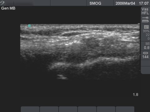
The area of injury is marked while taking into account the clinical exam, and data from imaging studies such as MRI and radiographs. It is recommended to use Dynamic musculoskeletal ultrasound with a transducer of 613 Hz in an effort to more accurately Localize the PRP injection. Under sterile conditions, the patient receives a PRP injection with or without approximately 1 cc of 1% Lidocaine and 1 cc of 0.25% Marcaine directly into the area of injury. Calcium chloride and Thrombin may be added to provide a Gel matrix for the PRP to adhere to, potentially maximizing the benefit in the case of a Joint space. We recommend using a Peppering technique spreading in a Clock-like manner to achieve a more expansive zone of delivery. The patient is observed in a supine position for 15–20 min afterwards, and is then discharged home. Patients typically experience minimal to moderate discomfort following the injection which may last for up to 1 week. They are instructed to ice the injected area if needed for pain control in addition to elevation of the limb and modification of activity as tolerated. We recommend Acetaminophen as the optimal analgesic, or Vicodin for break through pain, and dissuade the use of NSAID’s in the early post-injection period (Fig. 6).


Fig. 6

Musculoskeletal ultrasound, common extensor tendinosis


Safety

Any concerns of immunogenic reactions or disease transfer are eliminated because PRP is prepared from autologous blood. No studies have documented that PRP promotes hyperplasia, carcinogenesis, or tumor growth. Growth factors act on cell membranes rather than on the cell nucleus and activate normal gene expression [7]. Growth Factors are not mutagenic and naturally act through gene regulation and normal wound healing feed-back control mechanisms [6]. Relative contraindications include the presence of a Tumor, Metastatic disease, active Infections, or Platelet count < 10 5/ulHgb < 10 g/dl. Pregnancy or active Breastfeeding are contraindications. Patients with an Allergy to Bupivicaine (Marcaine) should not receive a local anesthetic with these substances.

The patients should be informed of the possibility of temporary worsening symptoms after the injection. This is likely due to the stimulation of the body’s natural response to Inflammatory mediators. Although adverse effects are uncommon, as with any injection there is a possibility of infection, no relief of symptoms, and neurovascular injury. Scar tissue formation and Calcification at the injection site are also remote risks.

An Allergic reaction or Local toxicity to Bupivacaine HCL or Lidocaine, although uncommon could trigger an adverse reaction. Additionally, when used in Surgical applications for grafting or with Intra-articular injections, PRP may be combined with Calcium chloride and bovine Thrombin to form a gel matrix. This bovine thrombin which is used to activate PRP, in the past has been associated with life threatening coagulopathies as a result of antibodies to clotting factors V, XI, and thrombin [7, 26]. However, since 1997 production has eliminated contamination of bovine thrombin with bovine factor Va. Prior to 1997, Va levels were 50 mg/ml and now are <0.2 mg/ml with no further reports of complications [6].

Charlie 發表在 痞客邦 留言(0) 人氣()

12178_2008_9032_Fig3_HTML  12178_2008_9032_Fig4_HTML  12178_2008_9032_Fig5_HTML  

Platelet rich plasma Injection grafts for musculoskeletal injuriesa review3.

PRP preparation

Various blood separation devices have differing preparation steps essentially accomplishing similar goals. The Biomet Biologics GPS III system is described here for simplicity. About 3060 ml of venous blood is drawn with aseptic technique from the anticubital vein. An 18 or 19 g butterfly needle is advised, in efforts of avoiding irritation and trauma to the platelets which are in a resting state. The blood is then placed in an FDA approved device and Centrifuged for 15 min at 3,200 rpm (Fig. 3). Afterward, the blood

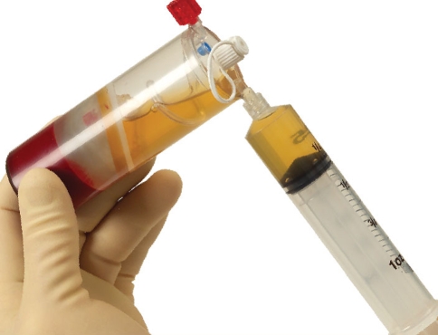
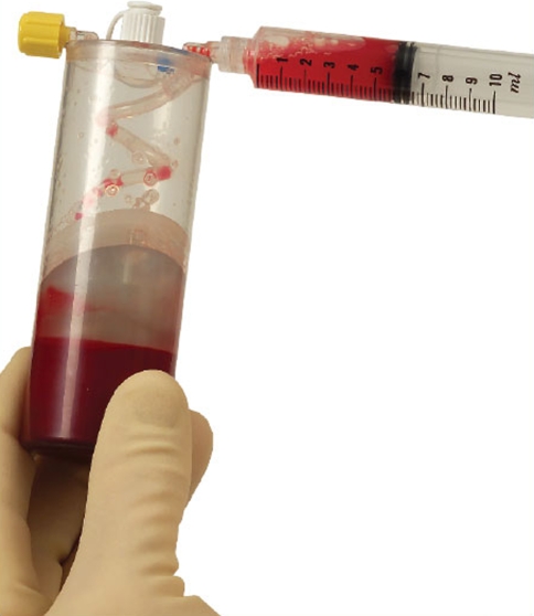
is separated into platelet poor plasma (PPP), RBC, and PRP. Next the PPP is extracted through a special port and discarded from the device (Fig. 4). While the PRP is in a vacuumed space, the device is shaken for 30 s to re-suspend the platelets. Afterwards the PRP is withdrawn (Fig. 5). Depending on the initial blood draw, there is approximately 3 or 6 cc of PRP available.


Fig. 3

GPS III system and centrifuge

Fig. 4

GPS III system, withdrawing of platelet poor plasma to be discarded

Fig. 5

GPS III withdrawing of platelet rich plasma for injection/graft

Charlie 發表在 痞客邦 留言(0) 人氣()

12178_2008_9032_Fig2_HTML  

Platelet rich plasma Injection grafts for musculoskeletal injuriesa review2.

TGFbeta is active during inflammation, and influences the regulation of cellular migration and proliferation; stimulate cell replication, and fibronectin binding interactions [23] (Fig. 2). VEGF is produced at its Highest levels only After the inflammatory phase, and is a potent stimulator of angiogenesis. Anitua et al. showed that in vitro VEGF and Hepatocyte Growth Factor (HGF) considerably increased following exposure to the pool of released growth factors; suggesting they accelerate Tendon cell proliferation and stimulate type I Collagen synthesis [11]. PDGF is produced following tendon damage and helps stimulate the production of other growth factors and has roles in tissue remodeling. PDGF promotes Mesenchymal stem cell replication, Osteoid production, Endothelial cell replication, and Collagen synthesis. It is likely the First growth factor present in a wound and starts connective tissue healing by promoting collagen and protein synthesis [7]. However, a recent animal study by Ranly et al. suggests that PDGF may actually inhibit bone growth [24].


Fig. 2

Active platelets


In vitro and in vivo studies have shown that bFGF is both a powerful Stimulator of Angiogenesis and a Regulator of Cellular migration and proliferation [23]. IGF-I is highly expressed during the early inflammatory phase in a number of animal tendon healing models, and likely assists in the proliferation and migration of Fibroblasts and to increase Collagen production [23]. However, a laboratory analysis of human PRP samples demonstrated increased concentrations of PDGF, TGFbeta, VEGF, and EGF, while not showing an increase in IGF-1 [25]. EGF effects are limited to Basal cells of Skin and Mucous membrane while inducing cell migration and replication.

Charlie 發表在 痞客邦 留言(0) 人氣()

12178_2008_9032_Fig1_HTML  

Platelet rich plasma Injection grafts for musculoskeletal injuriesa review1.

Steven Sampson,1 Michael Gerhardt,2 and Bert Mandelbaum2

1The Orthobiologic Institute (TOBI), Santa Monica, CA USA

2Santa Monica Orthopaedic Group, Santa Monica, CA USA

Steven Sampson, Email: drsampson@orthohealing.com.

Corresponding author.

Curr Rev Musculoskelet Med. 2008 December; 1(3-4): 165–174.

Published online 2008 July 16.

doi : 10.1007/s12178-008-9032-5

PMCID : PMC2682411

http://www.ncbi.nlm.nih.gov/pmc/articles/PMC2682411/


Abstract

In Europe and the United States, there is an increasing prevalence of the use of autologous blood products to facilitate healing in a variety of applications. Recently, we have learned more about specific growth factors, which play a crucial role in the healing process. With that knowledge there is abundant enthusiasm in the application of concentrated platelets, which release a supra-maximal quantity of these growth factors to stimulate recovery in non-healing injuries. For 20 years, the application of autologous PRP has been safely used and documented in many fields including; orthopedics, sports medicine, dentistry, ENT, neurosurgery, ophthalmology, urology, wound healing, cosmetic, cardiothoracic, and maxillofacial surgery. This article introduces the reader to PRP therapy and reviews the current literature on this emerging treatment modality. In summary, PRP provides a promising alternative to surgery by promoting safe and natural healing. However, there are few controlled trials, and mostly anecdotal or case reports. Additionally the sample sizes are frequently small, limiting the generalization of the findings. Recently, there is emerging literature on the beneficial effects of PRP for chronic non-healing tendon injuries including Lateral epicondylitis and Plantar fasciitis and Cartilage degeneration (Mishra and Pavelko, The American Journal of Sports Medicine 10(10):1–5, 2006; Barrett and Erredge, Podiatry Today 17:37–42, 2004). However, as clinical use increases, more controlled studies are needed to further understand this treatment.


Keywords : Platelet rich plasma, Injection, Growth factors, Tendon injury, Autologous blood, Musculoskeletal injuries, Chondropenia, Knee osteoarthritis


Introduction

In Europe, and more recently in the United States, an increased trend has emerged in the use of autologous blood products in an effort to facilitate healing in a variety of applications. In recent years, scientific research and technology has provided a new perspective on understanding the wound healing process. Initially platelets were thought to act exclusively with clotting. However, we have learned that platelets also release many bioactive proteins responsible for attracting Macrophages, Mesenchymal stem cells, and Osteoblasts which not only promotes removal of necrotic tissue, but also enhances tissue regeneration and healing.

Based on this principle platelets are introduced to stimulate a Supra-physiologic release of Growth factors in an attempt to Jump start healing in chronic injuries. The current literature reveals a paucity of randomized clinical trials. The existing literature is filled with mostly anecdotal reports or case series, which typically have small sample sizes and few control groups [1, 2]. A large multi-center trial is currently underway providing a more objective understanding of Platelet Rich Plasma (PRP) use in chronic epicondylitis.

According to the World Health Organization (WHO), musculoskeletal injuries are the most common cause of severe long-term pain and physical disability, and affect hundreds of millions of people around the world [3]. In fact, the years 2000–2010 have been termed “the decade of bone and joint” as a global initiative to promote further research on prevention, diagnosis, and treatment [3, 4]. Soft tissue injuries including Tendon and Ligament trauma represent 45% of all musculoskeletal injuries in the USA [4, 5]. The continued popularity of sporting activities has brought with it an epidemic of musculoskeletal disorders focusing attention on tendons. Additionally, modern imaging techniques including magnetic resonance imaging and musculoskeletal ultrasound have provided clinicians with further knowledge of these injuries.


Blood components

Blood contains plasma, red blood cells (RBC), white blood cells (WBC), and Platelets. Plasma is the liquid component of blood, made mostly of water and acts as a transporter for cells. Plasma also contains Fibrinogen, a protein that acts like a Net and Catches platelets at a wound site to form a clot. RBC helps pick up Oxygen from the lungs and delivers it to other body cells, while removing carbon dioxide. WBC fights infection, kills germs, and carries off dead blood cells. Platelets are responsible for Hemostasis, Construction of new connective tissue, and Revascularization. Typically a blood specimen contains 93% RBC, 6% Platelets, and 1% WBC [6]. The rationale for PRP benefit lies in reversing the blood ratio by decreasing RBC to 5%, which are less useful in the healing process, and increasing Platelets to 94% to stimulate recovery.


Platelets

Platelets are small discoid blood cells made in bone marrow with a lifespan of 7–10 days. Inside the platelets are many intracellular structures containing glycogen, lysosomes, and two types of granules. The alpha granules contain the Clotting and Growth factors that are eventually released in the healing process. Normally at the resting state, platelets require a trigger to activate and become a participant in wound healing and hemostasis [7]. Upon activation by Thrombin, the platelets morph into different shapes and develop branches, called Pseudo-pods that spread over injured tissue. This process is termed Aggregation. Eventually the granules contained within platelets release the Growth factors, which stimulate the Inflammatory cascade and Healing [7].


PRP

Platelet Rich Plasma is defined as a volume of the plasma fraction of autologous blood having a platelet concentration above baseline [8, 9]. Normal platelet concentration is 200,000 Platelets/ul. Studies have shown that clinical efficacy can be expected with a minimum increase of 4× this baseline (1million Platelets/ul) [6]. Slight variability exists in the ability to concentrate platelets, largely depending on the manufacturer’s equipment. However, it has not been studied if too great an increased platelet concentration would have paradoxical effects.

The use of autologous PRP was first used in 1987 by Ferrari et al. [10] following an open heart surgery, to avoid excessive transfusion of homologous blood products. Since that time, the application of autologous PRP has been safely used and documented in many fields including; orthopedics, sports medicine, dentistry, ENT, neurosurgery, ophthalmology, urology, and wound healing; as well as cosmetic, cardiothoracic, and maxillofacial surgery. Studies suggest that PRP can affect Inflammation, Post-operative blood loss, Infection, narcotic requirements, Osteogenesis, Wound, and Soft tissue healing.

In addition to Local hemostasis at sites of vascular injury, platelets contain an abundance of Growth factors and Cytokines that are pivotal in Soft tissue healing and Bone mineralization [4]. An increased awareness of platelets and their role in the healing process has lead to the concept of therapeutic applications.


Tendons

PRP is increasingly used in treatment of Chronic non-healing Tendon injuries including the elbow, patella, and the achilles among others. As a result of mechanical factors, tendons are vulnerable to injury and stubborn to heal. Tendons are made of specialized cells including Tenocytes, water, and fibrous Collagen proteins. Millions of these collagen proteins weave together to form a durable strand of flexible tissue to make up a tendon. They naturally anchor to the bone and form a resilient Mineralized connection. Tendons also bear the responsibility of transferring a great deal of force, and as a result are susceptible to injury when they are overwhelmed. With Repetitive overuse, Collagen fibers in the tendon may form Micro tears, leading to what is called Tendonitis; or more appropriately Tendinosis or Tendinopathy. The injured tendons heal by Scarring which adversely affects function and increases risk of re-injury. Furthermore, tendons heal at a Slow rate compared with other connective tissues, secondary to Poor vascularization [11–13]. Histologic samples from chronic cases indicate that there is Not an inflammatory response, But rather a limitation of the normal tendon repair system with a fibroblastic and a vascular response called, Angiofibroblastic degeneration [1, 14, 15]. Given the inherent nature of the tendon, new treatment options including Dry needling, Prolotherapy, and Extracorporeal shockwave therapy are aimed at embracing Inflammation rather than suppressing it.

Traditional therapies to treat these conditions do not alter the tendon’s inherent poor healing properties and involve long-term palliative care [16, 17]. A recent meta-analysis of 23 randomized controlled studies on physical therapy treatment for epicondylitis, concluded that there is insufficient supportive evidence of improved outcomes [1, 18]. Corticosteroids are commonly injected, however studies suggest Adverse side effects including atrophy and permanent adverse structural changes in the tendon [14]. Medications including NSAIDs, while commonly used for tendinopathies, carry significant long-term risks including bleeding ulcers and kidney damage. Thus, organically based strategies to promote healing while facilitating the release of one’s own natural growth factors is attracting interest.


Growth factors

It is widely accepted that Growth factors play a central role in the Healing process and tissue Regeneration [4, 19]. This conclusion has lead to significant research efforts examining varying growth factors and their role in repair of tissues [4, 20]. However, there are conflicting reports in the literature regarding potential benefits. Although some authors have reported improved bone formation and tissue healing with PRP, others have had less success [4, 21, 22]. These varying results are likely attributed to the need for additional Standardized PRP protocols, preparations, and techniques. There are a variety of commercially FDA approved kits available with variable platelet concentrations, clot activators, and leukocyte counts which could theoretically affect the data.

Alpha granules are storage units within platelets, which contain pre-packaged Growth factors in an inactive form (Fig. 1). The main growth factors contained in these granules are transforming growth

factor beta (TGFbeta), vascular endothelial growth factor (VEGF) platelet-derived growth factor (PDGF), and epithelial growth factor (EGF) (Table 1). The granules also contain Vitronectin, a cell adhesion molecule which helps with osseointegration and osseoconduction.


Fig. 1

Inactive platelets

Table 1

Growth factor chart [Printed with permission from: Eppley BL, Woodell JE, Higgins J. Platelet quantification and growth factor analysis from platelet-rich plasma: implications for wound healing. Plast Reconstr Surg. 2004 November;114(6):1502–8]

Platelet-derived growth factor (PDGF)

Stimulates cell replication

Promotes angiogenesis

Promotes epithelialization

Promotes granulation tissue formation

Transforming growth factor (TGF)

Promotes formation of extracellular matrix

Regulates bone cell metabolism

Vascular endothelial growth factor (VEGF)r

Promotes angiogenesis

Epidermal growth factor (EGF)

Promotes cell differentiation and stimulates re-epithelialisation, angiogenesis and collagenase activity

Fibroblast growth factor (FGF)

Promotes proliferation of endothelial cells and fibroblasts

Stimulates angiogenesis

Charlie 發表在 痞客邦 留言(0) 人氣()

1-s2.0-S1079210405005913-gr1

 1-s2.0-S1079210405005913-gr2  1-s2.0-S1079210405005913-gr3

Platelet-rich fibrinPRFa second-generation platelet concentrate. Part Vhistologic evaluations of PRF effects on bone allograft maturation in sinus lift.

Choukroun J, Diss A, Simonpieri A, Girard MO, Schoeffler C, Dohan SL, Dohan AJ, Mouhyi J, Dohan DM.

Oral Surg Oral Med Oral Pathol Oral Radiol Endod. 2006 Mar;101(3):299-303.

Oral Surgery, Oral Medicine, Oral Pathology, Oral Radiology, and Endodontology Volume 101, Issue 3, March 2006, Pages 299–303


Source

Pain Clinic Center, Nice, France.

http://www.sciencedirect.com/science/article/pii/S1079210405005913


Abstract

OBJECTIVE:

Platelet-rich fibrin (PRF) belongs to a new generation of platelet concentrates, with simplified processing and without biochemical blood handling. The use of platelet gel to improve bone regeneration is a recent technique in implantology. However, the biologic properties and real effects of such products remain controversial. In this article, we therefore attempt to evaluate the potential of PRF in combination with freeze-dried bone allograft (FDBA) (Phoenix; TBF, France) to enhance bone regeneration in sinus floor elevation.


STUDY DESIGN:

Nine sinus floor augmentations were performed. In 6 sites, PRF was added to FDBA particles (test group), and in 3 sites FDBA without PRF was used (control group). Four months later for the test group and 8 months later for the control group, bone specimens were harvested from the augmented region during the implant insertion procedure. These specimens were treated for histologic analysis.


RESULTS:

Histologic evaluations reveal the presence of residual bone surrounded by newly formed bone and connective tissue. After 4 months of healing time, histologic maturation of the test group appears to be identical to that of the control group after a period of 8 months. Moreover, the quantities of newly formed bone were equivalent between the 2 protocols.


CONCLUSIONS:

Sinus floor augmentation with FDBA and PRF leads to a reduction of healing time prior to implant placement. From a histologic point of view, this healing time could be reduced to 4 months, but large-scale studies are still necessary to validate these first results.


Fig. 1. Preliminary analyses highlight mineralized trabecular bone rich in osteocytes which appear green (A and B) or blue (C and D) according to the staining. Osteoïd borders are stained in red (B and D) and are in contact with dense cellular osteoblast fronts. The richness of osteoïd tissue is evidence of important turnover in both types of samples (test and control).

Fig. 2. Mean histomorphometric analysis of bone samples from 3 sinus floor augmentations after a healing period of 8 months (control group: FDBA alone).

Fig. 3. Mean histomorphometric analysis of bone samples from 6 sinus floor augmentation after a healing period of 4 months (test group: FDBA+PRF).


Charlie 發表在 痞客邦 留言(0) 人氣()

1-s2.0-S1079210405005895-gr1  1-s2.0-S1079210405005895-gr2  

Platelet-rich fibrinPRFa second-generation platelet concentrate. Part IVClinical effects on tissue healing.

Choukroun J, Diss A, Simonpieri A, Girard MO, Schoeffler C, Dohan SL, Dohan AJ, Mouhyi J, Dohan DM.

Oral Surg Oral Med Oral Pathol Oral Radiol Endod. 2006 Mar;101(3):e56-60.

Oral Surgery, Oral Medicine, Oral Pathology, Oral Radiology, and Endodontology Volume 101, Issue 3, March 2006, Pages e56–e60


Source

Pain Clinic Center, Nice, France.

http://www.sciencedirect.com/science/article/pii/S1079210405005895


Abstract

Platelet-rich fibrin (PRF) belongs to a new generation of platelet concentrates, with simplified processing and without biochemical blood handling. In this fourth article, investigation is made into the previously evaluated biology of PRF with the first established clinical results, to determine the potential fields of application for this biomaterial. The reasoning is structured around 4 fundamental events of cicatrization, namely, Angiogenesis, Immune control, Circulating stem cells trapping, and Wound-covering epithelialization. All of the known clinical applications of PRF highlight an accelerated tissue cicatrization due to the development of effective Neovascularization, accelerated wound Closing with fast cicatricial tissue Remodelling, and nearly total Absence of infectious events. This initial research therefore makes it possible to plan several future PRF applications, including plastic and bone surgery, provided that the real effects are evaluated both impartially and rigorously.


Fig. 1. Tooth extraction and osseous filling in a case of terminal periodontitis of wide sites (A and B) are delicate interventions because of the difficulty in obtaining soft tissue coverage on the surface of the osseous injury. Sockets are filled with Phoenix allogenic bone (TBF, France), (C). The use of PRF as cover membranes (D and E) permits a rapid epithelialization of the surface of the site, neutralizing the infectious phenomena. Forty-eight hours postoperative, wound is totally closed and sutures are removed (F).

Fig. 2. During massive cystic ablation of the maxillary (A and B), residual cavity is filled with PRF (C). Two and a half months later, the osseous defect is replaced by a dense and cortical bone (D) instead of the average 10 months naturally. The use of PRF allows acceleration of the physiologic phenomena.

Charlie 發表在 痞客邦 留言(0) 人氣()

1-s2.0-S1079210405005883-gr1  

1-s2.0-S1079210405005883-gr2  1-s2.0-S1079210405005883-gr3  1-s2.0-S1079210405005883-gr4  1-s2.0-S1079210405005883-gr5  1-s2.0-S1079210405005883-gr6  

Platelet-rich fibrinPRFa second-generation platelet concentrate. Part IIILeucocyte activationa new feature for platelet concentrates

Dohan DM, Choukroun J, Diss A, Dohan SL, Dohan AJ, Mouhyi J, Gogly B.

Oral Surg Oral Med Oral Pathol Oral Radiol Endod. 2006 Mar;101(3):e51-5.

Oral Surgery, Oral Medicine, Oral Pathology, Oral Radiology, and Endodontology Volume 101, Issue 3, March 2006, Pages e51–e55


Source

Biophysics Laboratory, Faculty of Dental Surgery, University of Paris V, Paris, France. drdohand@hotmail.com

http://www.sciencedirect.com/science/article/pii/S1079210405005883


Abstract

Platelet-rich fibrin (PRF) belongs to a new generation of platelet concentrates, with simplified processing and without biochemical blood handling. In this third article, we investigate the immune features of this biomaterial. During PRF processing, Leucocytes could also secrete Cytokines in reaction to the Hemostatic and Inflammatory phenomena artificially induced in the centrifuged tube. We therefore undertook to quantify 5 significant cell mediators within platelet poor plasma supernatant and PRF clot exudate serum : 3 proinflammatory cytokines (IL-1beta, IL-6, and TNF-alpha), an antiinflammatory cytokine (IL-4), and a key growth promoter of angiogenesis (VEGF). Our data are correlated with that obtained in plasma (nonactivated blood) and in sera (activated blood). These initial analyses revealed that PRF could be an immune regulation node with inflammation retrocontrol abilities. This concept could explain the reduction of postoperative infections when PRF is used as surgical additive.


Fig. 1. Schematic representation of the 3 centrifugation strata obtained after PRF processing according to the official Process protocol.

Fig. 2. IL-1β ELISA Quantifications.

Fig. 3. IL-6 ELISA Quantifications.

Fig. 4. TNF-α ELISA Quantifications.

Fig. 5. IL-4 ELISA Quantifications.

Fig. 6. VEGF ELISA Quantifications.

Charlie 發表在 痞客邦 留言(0) 人氣()

1-s2.0-S1079210405005871-gr1   1-s2.0-S1079210405005871-gr2

 1-s2.0-S1079210405005871-gr3  1-s2.0-S1079210405005871-gr4

 1-s2.0-S1079210405005871-gr5  1-s2.0-S1079210405005871-gr6  1-s2.0-S1079210405005871-gr7

 1-s2.0-S1079210405005871-gr8  1-s2.0-S1079210405005871-gr9  1-s2.0-S1079210405005871-gr10  

Platelet-rich fibrinPRF):a second-generation platelet concentrate. Part IIPlatelet-related biologic features.

Dohan DM, Choukroun J, Diss A, Dohan SL, Dohan AJ, Mouhyi J, Gogly B.

Oral Surg Oral Med Oral Pathol Oral Radiol Endod. 2006 Mar;101(3):e45-50. Epub 2006 Jan 10.

Oral Surgery, Oral Medicine, Oral Pathology, Oral Radiology, and Endodontology Volume 101, Issue 3, March 2006, Pages e45–e50


Source

Biophysics Laboratory, Faculty of Dental Surgery, University of Paris V, Paris, France. drdohand@hotmail.com

http://www.sciencedirect.com/science/article/pii/S1079210405005871


Abstract

Platelet-rich fibrin (PRF) belongs to a new generation of platelet concentrates, with simplified processing and without biochemical blood handling. In this second article, we investigate the platelet-associated features of this biomaterial. During PRF processing by centrifugation, platelets are activated and their massive degranulation implies a very significant cytokine release. Concentrated platelet-rich plasma platelet cytokines have already been quantified in many technologic configurations. To carry out a comparative study, we therefore undertook to quantify PDGF-BB, TGFbeta-1, and IGF-I within PPPplatelet-poor plasmasupernatant and PRF clot exudate serum. These initial analyses revealed that slow fibrin polymerization during PRF processing leads to the intrinsic incorporation of platelet cytokines and glycanic chains in the fibrin meshes. This result would imply that PRF, unlike the other platelet concentrates, would be able to progressively release Cytokines during Fibrin matrix remodeling; such a mechanism might explain the clinically observed healing properties of PRF.


Fig. 1. The lower part of the PRF fibrin matrix is occupied by whitish streaks looking like cell fragment aggregates on histological sections. These are the platelet accumulations and constitute a “buffy coat” (A). But there is no platelet or any other cellular body in the upper part of the PRF fibrin clot (B). Hemalun-eosin staining, 52×.

Fig. 2. The PRF fibrin clot obtained according to the Process protocol is divided into 3 parts: a red thrombus in contact with the red blood corpuscle base, an acellular fibrin gel, and a network of buffy columns corresponding to platelet accumulation.

Fig. 3. Glycanic chain distribution within PRF fibrin clot (pH 1 alcian blue staining, 52×).

Fig. 4. Schematic representation of the 3 centrifugation strata obtained after PRF processing according to Process official protocol.

Fig. 5. PDGF-BB ELISA quantifications.

Fig. 6. TGFβ-1 ELISA quantifications.

Fig. 7. IGF-1 ELISA quantifications.

Fig. 8. Theoretical computer modeling of a fibrin network resulting from fibrin glue polymerization. Note that in adhesives such as Tisseel, fibronectin is trapped in the matricial meshes (not represented here) (D-TEP v1.3).

Fig. 9. Theoretical computer modeling of a fibrin network resulting from a cPRP polymerization. The activated platelets are trapped in the fibrin meshes and release a significant quantity of cytokines extrinsically retained in the fibrin architecture (D-TEP v1.3; scales not respected). (1) Platelet trapped in the fibrin gel. (2) Platelet cytokine in solution (extrinsic).

Fig. 10. Theoretical computer modeling of a PRF clot. Note the presence of structural glycoproteins (fibronectin) and extrinsic cytokines (in solution) enmeshed in the fibrin matrix. The PRF slow polymerization process would also allow the intrinsic retaining of glycanic chains and cytokines within fibrin polymers. PRF would be thus very close to a natural fibrin thrombus (D-TEP v1.3; scales not respected). (1) Cytokine intrinsically retained within fibrin fibrillae. (2) Platelet cytokine in solution (extrinsically associated with fibrin polymers). (3) Fibrin-associated glycanic chains. (4) Circulating glycoproteins (fibronectin). (5) Fibrin fibrilla associated with glycanic chains and intrinsic cytokines.

Charlie 發表在 痞客邦 留言(0) 人氣()