目前分類:Cell 細胞 (179)

瀏覽方式: 標題列表 簡短摘要

152d3ce0a3c864152d3ce1f7a9c1152d3ce2c1af19152d3ce3a70d82152d3ce32a3f89152d3ce3fd9b36152d3ce107a077152d3ce265e7f8152d3ce459160f  

Apoptosis
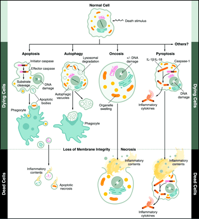
Apoptosis is the process of Programmed cell deathPCDthat may occur in multicellular organisms.
Biochemical events lead to characteristic cell changes (morphology) and death. These changes include blebbing, cell shrinkage, nuclear fragmentation, chromatin condensation, and chromosomal DNA fragmentation.
In contrast to necrosis, which is a form of traumatic cell death that results from acute cellular injury, apoptosis generally confers Advantages during an organism's life cycle. For example, the separation of fingers and toes in a developing human embryo occurs because cells between the digits apoptose. Unlike necrosis, apoptosis produces cell fragments called Apoptotic bodies that phagocytic cells are able to engulf and quickly remove before the contents of the cell can spill out onto surrounding cells and cause damage.
Between 50 and 70 billion cells die each day due to apoptosis in the average human adult. For an average child between the ages of 8 and 14, approximately 20 billion to 30 billion cells die a day.
Research in and around apoptosis has increased substantially since the early 1990s. In addition to its importance as a biological phenomenon, Defective apoptotic processes have been implicated in an extensive variety of diseases. Excessive apoptosis causes atrophy, whereas an insufficient amount results in Uncontrolled cell proliferation, such as cancer.
 
Summary of the different Morphologies, Mechanisms and Outcomes of the 3 forms of cell deathApopotosis, Pyroptosis and Necrosis

  Characteristics Apoptosis Pyroptosis Necrosis
Morphology             
  Cell lysis X O O
  Cell swelling X O O
  Pore formation X O O
  Membrane blebbing O X X
  DNA fragmentation O O O
Mechanism        
  Caspase-1 X O X
  Caspase-3 O X X
  Cytochome-c release O X X
Outcome        
  Inflammation X O O
  Programmed cell death O O X

Charlie 發表在 痞客邦 留言(0) 人氣()

1-s2.0-S1521690X1200067X-gr1badi041mtdiscovery_medicine_clifford_j_rosen_no_58_figure_1fendo-02-00118-g001nrendo.2010.20-f3nrendo.2010.64-f2Stemcells1_0VisualAbstractYin-et-al300wcredit  

棕色脂肪能減重 吃蘋果草莓強效

作者:華人健康網記者黃曼瑩/台北報導 | 華人健康網 – 2014112


許多女性步入中年後,就不禁經常望著日漸隆起的小腹大嘆:「瘦不了」,為什麼「中年容易發福」?原來是「棕色脂肪」在作怪,根據日本一項研究發現,隨著年紀增長,人體內的「棕色脂肪」數量減少,因較難燃燒體內不良脂肪,於是使肥胖問題更加嚴重。減重專家建議,多攝取富含熊果酸的水果,有助於增加棕色脂肪,達到降低體內熱量的效果。

什麼是棕色脂肪?人體脂肪細胞有棕色和白色兩種脂肪。棕色脂肪負責分解引發肥胖的脂肪,將白色脂肪轉化成二氧化碳、水和熱量,可以說是「天生減肥專家」。棕色脂肪在嬰兒時期十分充足,隨著年齡增長而減少,大多數成人只有在上背和脖子等少部份區域存在棕色脂肪。

至於白色脂肪則是會在全身作為能量備份儲存,以作為各餐之間消耗所需;但當人們開始大量將毒素、加工食品和無用卡路里塞進肚,白色脂肪的累積超過負荷,便成為一種負擔

根據發表於新英格蘭雜誌(New England Journal of Medicine)的報告指出,在不同溫度下,棕色脂肪組織的活性不同,在低溫時,棕色脂肪組織會特別被活化。也就是說,隨著運動和接觸冷空氣的時間增加,棕色脂肪儲存也會增加


提升棕色脂肪第1招:洗冷水浴

新英格蘭雜誌的研究顯示,處於1619度的受試者,其棕色脂肪組織活化的程度明顯高於處於常溫下者,因此,想提升棕色脂肪代謝並燃燒多餘的熱量,就要少吹暖氣,而建議洗冷水澡增進棕色脂肪代謝;不過最有效的方式還是在冷天運動,運動結合低溫,會同時激活棕色脂肪細胞並分解代謝的白色脂肪。


提升棕色脂肪第2招:補充熊果素食物

此外,根據愛荷華大學研究團隊的研究發現,蘋果皮中富含的熊果酸,可以增加肌肉及棕色脂肪量,使身體燃燒熱量,對抗肥胖。熊果素是一種由杜鵑花科植物熊果葉中萃取出的成份,在梨、小麥等植物中也有發現,熊果素也是相當有名的美白成分。

天然的熊果素,大多存在於一些漿果類的植物中,例如大家熟知的藍莓、草莓、蔓越莓等,同時也可以在小紅莓、梅子、西洋梨、羅勒、百里香、花椒葉和麝香草中找到。


減重關鍵密碼 規律運動與減少熱量

雖然棕色脂肪組織的量對於減重有益,但是,想要減重成功,唯有靠規律運動與減少熱量攝取最才重要,唯有多管齊下,才能達到健康減重的長久效果。

Charlie 發表在 痞客邦 留言(0) 人氣()

F2.mediumF6.largeF8.largeInflammasome_final1ni.2224-F1Pyroptosis_1Pyroptosis_mechanism  

Pyroptosis

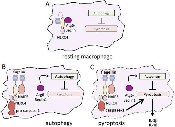
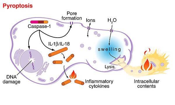
Pyroptosis is a form of Programmed cell death associated with Antimicrobial responses during Inflammation.

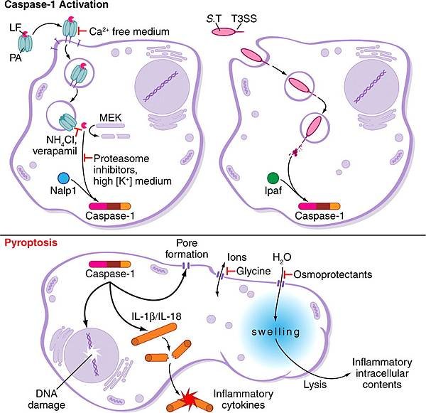
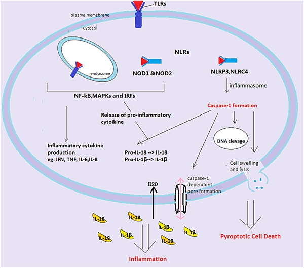
In contrast to apoptosis, pyroptosis requires the function of Caspase-1, and has been studied in the context of Salmonella-infected macrophages. The initiation of pyroptosis has been linked to the recognition of Flagellin components of Salmonella and Shigella species (and similar Pathogen-associated molecular patterns (PAMPs) in other microbial pathogens) by NOD-like receptors (NLRs). These receptors function like plasma membrane Toll-like receptors (TLRs), but recognise intracytoplasmic antigens rather than extracellular ones.

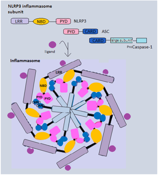
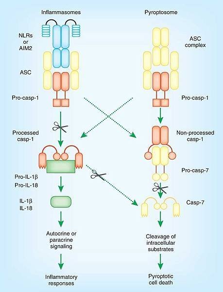
Recently, it was shown that Caspase-1 is activated during pyroptosis by a large supramolecular complex termed the Pyroptosome (also known as an Inflammasome).

Only one large PyroptosomeInflammasomeis formed in each macrophage, within minutes after infection. Biochemical and Mass Spectroscopic analysis revealed that this pyroptosome is largely composed of dimers of the adaptor protein ASC (apoptosis-associated speck protein containing a CARD or Caspase activation and recruitment domain).

Unlike apoptosis, Pyroptosis results in the release of Pathogen associated molecular patterns (PAMPs) and Cytokines that activate pro-inflammatory immune cell mediators.


Summary of the different Morphologies, Mechanisms and Outcomes of the 3 forms of cell deathApopotosis, Pyroptosis and Necrosis


Characteristics

Apoptosis

Pyroptosis

Necrosis

Morphology    


Cell lysis

X

O

O


Cell swelling

X

O

O


Pore formation

X

O

O


Membrane blebbing

O

X

X


DNA fragmentation

O

O

O

Mechanism


Caspase-1

X

O

X


Caspase-3

O

X

X


Cytochome-c release

O

X

X

Outcome


Inflammation

X

O

O


Programmed cell death

O

O

X


Charlie 發表在 痞客邦 留言(0) 人氣()

797px-Inflammasomes_activation_and_signaling_overviewinflammasomeinflammasome_complexInflammasome_pathwaysInflammasome-Therapeutic-ImplicationsMEDIUM_11882_2010_109_Fig1_HTMLni0210-105-F1nm0711-790-F1

Inflammasome

The inflammasome is a multiprotein oligomer consisting of Caspase 1, PYCARD, NALP and sometimes Caspase 5 (also known as caspase 11 or ICH-3). It is expressed in Myeloid cells and is a component of the innate immune system.

The exact composition of an inflammasome depends on the activator which initiates inflammasome assembly, e.g. dsRNA will trigger one inflammasome composition whereas asbestos will assemble a different variant.

The inflammasome promotes the maturation of inflammatory cytokines Interleukin 1β (IL-1β) and Interleukin 18 (IL-18).

The inflammasome is responsible for activation of inflammatory processes, and has been shown to induce cell pyroptosis, a process of programmed cell death distinct from apoptosis.

nri1997-f4  

Charlie 發表在 痞客邦 留言(0) 人氣()

file_237610ijbsv06p0796g05iPS_factsheet_diag_new_Dec2012.img_assist_custom-600x452ipsc-pathwayiPSCwhatcando_3karyotype-diagramnature10761-f2.2

news_8__782594814

nrm2466-f2Ring1Ring4ringchromosomeringswhat_is_sc

美國Gladstone Institute再生醫療新進展!iPSInduced pluripotent stem cells修復異常的環狀染色體Ring chromosomes

作者:NewTalk 新頭殼 | 新頭殼 – 2014113

新頭殼newtalk2014.01.13 鄭凱榕/編譯報導


美國格萊斯頓研究所(Gladstone Institute)等團隊,12日在英國科學雜誌《自然》(Nature)電子版上發表最新的研究結果。研究團隊從先天性擁有環狀異常染色體的病患皮膚,做成人工多能幹細胞,又稱為誘導性多功能幹細胞Induced pluripotent stem cells,簡稱iPS,可以修復異常染色體

根據日本共同通信社13日報導,研究團隊中的研究員林洋平(HAYASHI, Yohei)表示,「雖然修復的機制尚未清楚,具修復功能的iPS細胞使臟器和細胞發生變化,將來可能可以移植到患者本人身上,具備再生醫療應用的可能性。」

2012年諾貝爾生理醫學獎得主、京都大學的山中伸彌教授也參與了這項研究。

染色體在細胞之內,通常形狀為棒狀

環狀染色體則呈現兩端部分被切除後相連的環狀

根據我國衛生福利部官方網站上「遺傳小百科」中對於「染色體異常環狀染色體」的簡介,1條染色體上出現2個斷裂點時,若是這2個斷裂點發生融合就會形成環狀染色體Ring chromosomes不具有著絲點的環狀染色體在細胞分裂時會遺失,而具有著絲點的環狀染色體則會在細胞分裂時保存下來。在臨床上,1個環狀染色體表示存在有2個缺失


Charlie 發表在 痞客邦 留言(0) 人氣()

adi0211headipose_tissue_white_40x6ahima_white_adipose_tissuebadi041mt

bj430e001f01

bwfatijo2010183f1image3lago.fig1nri2174-f5White_adipose_distribution_in_the_body.

White adipose tissueWAT

White adipose tissue (WAT) or white fat is one of the two types of adipose tissue found in mammals. The other kind of adipose tissue is brown adipose tissue.

In healthy, non-overweight humans, white adipose tissue composes as much as 20% of the body weight in men and 25% of the body weight in women. Its cells contain a single large fat droplet, which forces the nucleus to be squeezed into a thin rim at the periphery. They have Receptors for Insulin, Growth hormones, Norepinephrine and Glucocorticoids.

White adipose tissue is used as a store of energy. Upon release of Insulin from the pancreas, white adipose cells' insulin receptors cause a Dephosphorylation cascade that lead to the Inactivation of hormone-sensitive lipase. It was previously thought that upon release of glucagon from the pancreas, Glucagon receptors cause a Phosphorylation cascade that Activates hormone-sensitive lipase, causing the breakdown of the stored fat to fatty acids, which are exported into the blood and bound to albumin, and glycerol, which is exported into the blood freely. There is actually no evidence at present that glucagon has any effect on white adipose tissue. Glucagon is now thought to act exclusively on the Liver to trigger Glycogenolysis and Gluconeogenesis. The trigger for this process in white adipose tissue is instead now thought to be Adrenocorticotropic hormone (ACTH), Adrenaline and Noradrenaline.

Fatty acids are taken up by Muscle and Cardiac tissue as a Fuel source, and Glycerol is taken up by the Liver for Gluconeogenesis.

White adipose tissue also acts as a Thermal insulator, helping to maintain body temperature.

The hormone Leptin is primarily manufactured in the adipocytes of white adipose tissue.


Charlie 發表在 痞客邦 留言(0) 人氣()

Brown-fat-to-fight-white-fatwhite-and-brown-fat

male and female

F4.largenrd770-f4nrendo.2010.64-f2

體內好脂肪罷工年紀愈大減重越難

作者:【記者蘇湘雲/綜合外電報導】 | 台灣新生報 – 201417


不少中年男女受肥胖所苦,常感覺減重成效不如年輕時來得好,日本最新研究發現,這可能是體內好脂肪失去工作效率,使得年紀越大,減重就越困難。

根據英國「每日郵報」報導,人體有兩種脂肪型態,分別為「好」棕色脂肪「壞」白色脂肪,棕色脂肪可以幫助提升新陳代謝、產生熱能。日本靜岡大學研究顯示,隨著一個人年齡越來越大,棕色脂肪運作就會越來越沒有效率、活化程度越來越差,導致減重變得越加困難。如果可以刺激棕色脂肪,讓棕色脂肪重新活躍起來,或許就能治療一些肥胖相關疾病。

一般人對白色脂肪比較熟悉,白色脂肪囤積於下腹、大腿等皮下組織部位,當吃太多,就會造成白色脂肪堆積。而棕色脂肪有助增加身體代謝、燃燒熱量,小寶寶肩胛骨周圍就帶有棕色脂肪,小寶寶出生後運用這些棕色脂肪維持體溫正常。過去一向認為,當一個人逐漸長大,因為身體不再需要,所以棕色脂肪便會消失。不過也有人發現,就算進入成年階段,身體還是帶有一些棕色脂肪,而女性體內的棕色脂肪含量比男性多

日本研究團隊近日在「美國實驗生物學聯會期刊」(FASEB Journal)發表研究指出,當一個人年紀逐漸增長,棕色脂肪產熱效能就會變得低落,這代表棕色脂肪活性變差,且燃燒的白色脂肪數量逐漸縮減

「美國實驗生物學聯會期刊」主編吉拉爾德威斯曼指出,年紀較大的人常會抱怨,無論如何認真運動、節食,減重成效還是比不上年輕人。從這項研究就能了解,可能是因為棕色脂肪逐漸罷工而引起這種現象。年紀較大的人若想成功減重,就要比年輕人更認真地運動、節制飲食


【記者蘇湘雲/台北報導】

每到冬天,大吃大喝的機會就會增加,有些人大吃大喝,吃進大魚大肉後,因為良心不安,就想喝點消脂茶,看看能不能減少膽固醇堆積。

振興醫院心臟血管內科主治醫師張鴻猷表示,坊間販售的所謂消脂茶產品成份多樣,有些是綠茶,有些含有纖維質,這些成份或許可以減少飲食油脂吸收,但對於降低膽固醇效果非常有限

張鴻猷醫師指出,這些保健食品、消脂茶中或許對減少中性脂肪、三酸甘油脂有點幫助,不過對減少壞膽固醇「低密度脂蛋白」(LDL)效果也是很有限,因此最重要的還是盡量節制飲食,少吃高膽固醇食物,多吃健康、天然食物,才是根本養生、保健之道。此外,身體有部份膽固醇來自肝臟,就算減少飲食中的膽固醇吸收,也不見得真能達到降血脂目標。

疾病方面,像腎病症候群、甲狀腺低下、糖尿病等慢性疾病也都可能造成膽固醇上升。

張鴻猷醫師說,像消脂茶、紅麴等食品降低膽固醇比率偏低,比較起來,藥物降膽固醇比率就比較高,降膽固醇幅度有時可達三、四成。很多人知道自己有高血脂問題,但卻不知道自己的血脂指數,也不了解治療目標為何,對於有心血管疾病、心肌梗塞病史患者,治療目標要設低一點,才能預防併發症發生。建議慢性病患三到六個月、健康成年一年追蹤一次。


Charlie 發表在 痞客邦 留言(0) 人氣()

幹細胞移植成功治癒玻璃娃娃

作者:【記者萬博超/台北報導】 | 台灣新生報 – 20131218


台灣、瑞典、新加坡子宮內幹細胞移植研究團隊昨(17)日發表在子宮內應用間質幹細胞移植,成功治療兩例先天成骨發育不全症胎兒(玻璃娃娃)的成果。

長庚醫院產科羊水幹細胞研究團隊負責人,同時也是台灣母胎醫學會理事長鄭博仁教授表示,參與的研究個案是一位第四型的先天成骨發育不全症胎兒,在26孕周經超音波檢查,及基因檢測確立診斷後,即安排在31孕周進行子宮內幹細胞移植,並於出生一歲半時進行第二次幹細胞移植,目前這個四歲的小女孩可以正常作息,行走,跳舞,未曾出現骨折現象,對於移植的間質幹細胞也未產生排斥現象,或發生副作用。

玻璃娃娃是一種先天性骨骼疾病,導致生長發育遲緩及反覆的骨折。

此一研究成果已刊登於昨日出刊的「幹細胞轉譯醫學」期刊(Stem Cell Translational Medicine)。

醫師指出,未來有機會進一步發展出子宮內胎兒疾病的治療模式,包含地中海型貧血,脊髓性肌肉萎縮症或是其他代謝性疾病。

鄭博仁進一步指出,續玻璃娃娃的治療之後,長庚正準備啟動一系列相關研究,例如人類羊水幹細胞的分離、純化、複製、貯存;間質幹細胞於胎兒脊髓性肌肉萎縮症治療之應用等。

長庚及英國倫敦大學學院、瑞典卡羅林斯卡學院,和新加坡國立大學合作,從事羊水間質幹細胞宮內治療的研究,以往被視為醫療廢棄物的羊水,未來都是可用於治療胎兒疾病的重要資源。

研究團隊之一的婦產科蕭勝文醫師指出,未來孕婦接受羊膜穿刺術將羊水取出進行胎兒染色體分析以及羊水基因晶片,多餘的羊水可進一步的在體外分離、純化、貯存,甚而可利用基因標記,加入治療性基因,針對部分胎兒先天異常,先前自存的羊水幹細胞可做為自體移植治療之用。

此外,健康的胎兒也可以將自己的幹細胞貯存起來,以備出生或成年後,做為器官修補或細胞移植的自體羊水幹細胞庫。

Charlie 發表在 痞客邦 留言(0) 人氣()

基因治療子宮內幹細胞移植玻璃娃娃快樂跑跳

作者:邱俐穎台北報導 | 中時電子報 – 20131218

中國時報【邱俐穎台北報導】


先天成骨發育不全症患者俗稱「玻璃娃娃」,因長不高、輕微碰撞容易骨折得名。瑞典、新加坡、台灣林口長庚醫院組成的國際團隊,透過子宮內幹細胞移植,成功治療一名台灣的玻璃娃娃,快5歲的江小妹能走、能跳,和正常孩童無異。

林口長庚產科主任鄭博仁指出,玻璃娃娃盛行率約2萬分之一,若以國內每年新生兒出生數為20萬推估,每一年約有10幾位玻璃娃娃誕生,目前只能透過定期施打刺激骨骼生長的藥物,控制疾病嚴重度。

鄭博仁說,這名孕婦懷孕26周經超音波、基因檢測發現胎兒患有第4型先天成骨發育不全症,胎兒只是在子宮內羊水翻滾,左右大腿、手臂卻都骨折。

由於台灣醫療法規仍未核准幹細胞進行胎內移植,這名孕婦後來赴新加坡,利用瑞典卡羅林斯卡醫學院提供的間質幹細胞,進行子宮內幹細胞移植,移植才2周,胎兒原本骨折、斷裂的左大腿骨竟神奇癒合,出生時僅剩右大腿局部骨折

不過,出生後江小妹生長發育緩慢、落後同齡孩童,因此在1歲半時再次赴新加坡經靜脈注射進行第2次幹細胞移植。目前快5歲的她可唱歌、跳舞、行動自如,至今未發生過骨折

鄭博仁說,子宮內進行幹細胞移植優點在於胎兒自身免疫系統尚未形成,移植後的細胞會被玻璃娃娃身體辨識為自我細胞,因此1歲半進行第2次幹細胞移植也不會產生排斥。

林口長庚婦產部主治醫師蕭勝文表示,子宮內幹細胞移植等於是開啟了基因治療另一扇窗。這項重要的治療成果已發表在《幹細胞轉譯醫學》期刊。

蕭勝文還說,長庚醫院著手進行羊水間質幹細胞子宮內治療研究,過往被視為醫療廢棄物的羊水,可望運用在治療地中海型貧血、脊髓性肌肉萎縮症等疾病,但目前仍在動物實驗階段。

幹細胞是人類最原始的細胞,具有分裂、增殖為各種人體器官、組織的能力,最原始的幹細胞則是胚胎幹細胞,分化為身體細胞的潛力最大。

至於間質幹細胞雖不像胚胎幹細胞原始,但仍具有分化為人體骨骼、肌肉、脂肪,甚至神經、血管等組織細胞的潛力。

Charlie 發表在 痞客邦 留言(0) 人氣()

子宮內幹細胞移植治癒玻璃娃娃

自由時報 – 20131218

〔自由時報記者洪素卿/台北報導〕

成功擺脫玻璃娃娃命運

幹細胞移植,在娘胎裡也能做!林口長庚醫院與瑞典、新加坡等國際醫療團隊合作進行子宮內幹細胞移植研究,直接將間質幹細胞注射到還在媽媽肚子裡的胎兒身體,成功治療了兩例俗稱「玻璃娃娃」的先天成骨發育不全症胎兒。

其中,台灣個案在媽媽肚子裡已骨折,但經由兩度幹細胞移植後,目前已四歲的這個小女孩,現在就如同其他同齡女孩一樣,可正常作息、行走、跳舞,也未再出現骨折現象。

國際醫療團隊昨召開記者會說明此研究,相關論文也發表於甫出刊的「幹細胞轉譯醫學」期刊(Stem Cell Translational Medicine)。參與此國際研究的是林口長庚醫院婦產部產科醫師鄭博仁、蕭勝文和小兒科醫師林如立。


26週胎兒已經出現骨折

鄭博仁指出,台灣個案的媽媽在懷孕22週產檢時,醫師從超音波影像看到胎兒大腿骨、手臂骨等長骨相對短,有異常跡象。到26週,胎兒已有骨折跡象。經基因診斷確認,胎兒罹患第四型的成骨不全症,亦即俗稱的玻璃娃娃。

當時家屬很掙扎,但經檢查發現,原來家族中不只一人罹患此症,但他們除了小時候常骨折、身高較矮外,其他並無異於一般人。加上長庚恰好有此一國際合作計畫,因此,經醫療團隊與家屬溝通後,決定到新加坡讓肚子裡的寶寶接受胎內幹細胞治療。


胎內治療寶寶不會排斥

蕭勝文指出,這項治療的特殊性是在「胎內治療」,也就是趁寶寶還在媽媽肚子裡的時候移植。這時候孩子對外來的間質幹細胞不會排斥。

研究團隊在胎兒31週大時,以胎內注射方式,直接將瑞典培養的間質幹細胞注入胎兒的肝靜脈。待胎兒38週大時,剖腹生產

小女孩19個月大時、醫療團隊為她進行第二次幹細胞移植,這次則是直接以靜脈注射方式、將幹細胞注入小女孩體內。鄭博仁指出,經由第一次胎內幹細胞移植治療後,胎兒骨折逐漸癒合,出生後也無骨折現象。但小女孩的生長曲線卻有落後跡象,可能是因胎內注射幹細胞漸失功效,因此又二度移植;不過,現在身高仍比同齡小孩嬌小一些。

鄭博仁說,小女孩的母親後來懷了第二胎,很早就接受了絨毛採樣基因篩檢,結果檢驗結果正常。

Charlie 發表在 痞客邦 留言(0) 人氣()

48014

子宮內幹細胞移植 玻璃娃娃能跳了自存羊水幹細胞 可做為自體移植

作者:華人健康網記者黃曼瑩/台北報導 | 華人健康網 – 20131217

跨國醫療團隊在子宮內應用間質幹細胞,成功移植且治療兩例先天成


國內參與國際幹細胞移植技術再突破!經由跨國包括瑞典、新加坡與台灣在內的醫療團隊,在子宮內應用間質幹細胞,成功移植且治療兩例先天成骨發育不全症胎兒(即玻璃娃娃)。值得注意的是,以往被視為醫療廢棄物的羊水,在這些研究群專家的眼裡,成為治療胎兒疾病的重要黃金!

台灣參與此一國際研究團隊的是林口長庚醫院婦產部產科鄭博仁主任、蕭勝文醫師和小兒科林如立醫師。長庚醫院參與的研究個案是一位第四型的先天成骨發育不全症胎兒,在26孕周經超音波檢查,及基因檢測確立診斷後,即安排31孕周進行子宮內幹細胞移植,並於出生一歲半時進行第二次幹細胞移植,目前這個4歲的可愛小女孩和一般女生一樣,跟正常小孩一樣,能走、能跳,並未曾出現骨折現象,對於移植的間質幹細胞也未曾產生排斥現象,或是發生副作用。

此一研究成果論文並已發表刊登於今日出刊的「幹細胞轉譯醫學」期刊(Stem Cell Translational Medicine)。


自存羊水幹細胞 可做為自體移植

蕭勝文醫師表示,未來,孕婦於懷孕1620周期間,接受羊膜穿刺術將羊水取出進行胎兒染色體分析以及羊水基因晶片,多餘的羊水可進一步的在體外分離、純化、貯存,甚而可利用基因標記,加入治療性基因。針對部份胎兒先天異常,先前自存的羊水幹細胞可做為自體移植治療之用

另一方面,健康的胎兒也可以將自己的幹細胞貯存起來,以備出生或成年後,做為器官修補或細胞移植的自體羊水幹細胞庫。

長庚醫院產科羊水幹細胞研究團隊負責人,同時也是台灣母胎醫學會理事長鄭博仁教授表示,利用這個技術,可以進一步發展出子宮內胎兒疾病的治療模式,包含地中海型貧血、脊髓性肌肉萎縮症,或是其他代謝性疾病。

玻璃娃娃治療之後,長庚正準備啟動一系列相關研究,例如人類羊水幹細胞的分離、純化、複製、貯存;間質幹細胞於胎兒脊髓性肌肉萎縮症治療之應用等;可預見未來在亞太地區的子宮內移植幹細胞研究及臨床應用,佔有重要一席地位。


【醫學小辭典】:

先天成骨發育不全症是一種先天性遺傳疾病,一共有四大類型,最常見的是第一型,其次是第四型、第三型和第二型。出現在男女的比例上大約相同,這種疾病會造成第一型膠原纖維缺陷,使骨骼忍受外力衝擊的能力較正常人差,即使是輕微的碰撞,也會造成嚴重的骨折,因此這類的病患被稱為「玻璃娃娃」或「玻璃骨」。成骨不全症的發生機率為萬分之一以下,被列為罕見疾病,在台灣,每2萬至3萬人就有1名玻璃娃娃。


Charlie 發表在 痞客邦 留言(0) 人氣()

nrc3565-f1  nrc3565-f2  nrc3565-f3  nrc3565-f4

基因療法將白血球T細胞轉成搜尋並摧毀癌細胞的剋星治療血癌患者成效佳 Gene-engineered T cells for Cancer therapy

中央社 – 2013128

(中央社華盛頓8日綜合外電報導)

醫療團隊透過基因治療技術,將患者的白血球轉化成搜尋並摧毀癌細胞的剋星,成為醫學界多年來,對抗白血病和其他種類血癌的最大進展之一。

醫師群將在美國血液學會年會,報告這項空前的成果。

美聯社報導,一些罹患1種白血病的患者數年前接受這種一次性的實驗性治療後,若干人至今沒有出現癌細胞復發的情況。如今,至少有6個研究團體對超過120名罹患多種血液及骨髓癌症的患者進行治療,成效驚人。

西雅圖華盛頓大學(University of Washington)血液疾病專家、也是美國血液學會主席的阿布科維茲(Janis Abkowitz)教授說,「這非常令人興奮」。他表示,「你能取出患者體內的細胞,並將它轉化成攻擊型細胞」。

在一項研究中,有27名罹患急性淋巴球性白血病(ALL)的病患,其中5名成年人及22名兒童中的19人在接受治療後,病情徹底緩解,這意味著在接受治療後,患者體內已無癌細胞存在,不過有幾人日後出現復發的情況。

這些別無選擇的重症患者,其中有些人曾嘗試過多次骨髓移植和多達10種的化學治療或其他治療。

賓夕法尼亞州菲利普斯堡(Philipsburg8歲女童懷特黑德(Emily Whitehead)原已是癌症末期病患,醫師都說她的重要器官將於數日後衰竭。她是接受基因療法的首位兒童,接受治療近兩年後,至今仍無癌症復發的跡象。

有關其他罹患骨髓癌、淋巴瘤和慢性淋巴性白血病(CLL)患者的治療結果,將在紐奧良(New Orleans)舉行的美國血液學會(American Society of Hematology)年會中披露。

醫師表示,這有望成為美國及全球核准的第一種癌症基因治療技術;截至目前,歐洲僅核准一種治療罕見新陳代謝疾病的基因療法。

這種治療方法是先將患者血液進行過濾,移除數以百萬計稱作T細胞的白血球細胞,接著在實驗室進行基因改造,以植入一種以癌細胞為攻擊目標的基因,並在3天內將T細胞移回患者體內。


Charlie 發表在 痞客邦 留言(0) 人氣()

uho_news_030207  

糖尿病患腳部易潰爛 血小板成治癒關鍵

優活健康網 – 20131122

(優活健康網記者陳承璋/綜合報導)

在我國糖尿病患者突破兩百萬人,且新增人數居高不下的情況下,若病患血糖又時常控制不好,則容易產生許多併發症,而四肢的神經微血管病變,就是最常見的糖尿病併發症之一,導致腳部保護的不佳,常因小傷口造成經年累月不易恢復的慢性潰瘍傷口。若患者本身腎功能又不好,更會造成傷口長年難以癒合。


中年男腳爛 分離血小板治癒

而台中就有一位51歲鍾先生,因兩腳底皆有嚴重潰傷,由於傷口照顧不佳,造成傷口呈現糜爛潰瘍,且分泌物不斷,傷口四週及鞋上都被分泌物所沾黏。因傷口已沿用舊時的治療方式治療時間已超過一年,一直沒有改善,才到中國醫藥大學附設醫院求助,轉診至整形外科進行治療。

整形外科醫師張家寧在了解患者病史及與患者討論過後,決定採用病患自行的周邊血液提煉富血小板之血清來治療,患者接受治療一次後,傷口面積就小於原來面積的70%,患者在接受兩次治療後痊癒。

張家寧解釋,目前醫學在對於即時治癒糖尿病患者的慢性潰瘍有一定的瓶頸,而這種新治療方式是在病人身上抽取周邊靜脈血,當場分離血小板,可以在注射時增加傷口旁組織的生長激素,進而喚來全身或附近的幹細胞來修復組織(回家效應Homing),才能夠讓傷口變小、逐漸乾燥而且縮到痊癒。

但張家寧強調,在接受治療時,仍須注意傷口是否乾淨,所以施行新療法前,需先施行清創手術,之後的12週,再開始此療法,2週為一輪迴治療,來解決傷口癒合問題

Charlie 發表在 痞客邦 留言(0) 人氣()

2900129fd4_ksc_20131120_101010  

糖尿病足潰爛活化幹細胞療法傷口癒合

寇世菁報導中廣新聞網 – 20131120


台中一名51歲男子,有糖尿病,長期洗腎,手腳動輒破皮受傷,一年來,雙腳腳底嚴重潰瘍,傷口糜爛,分泌物不斷,連鞋子都沾到黏黏的,始終好不了,輾轉到中國附醫。整形外科採用病患自己的周邊血液,提煉富血小板的血清治療,刺激幹細胞活化,治療一次,傷口面積小於原面積的百分之七十,兩次治療後痊癒。

中國附醫整型外科主任張家寧表示,糖尿病足不容易治癒,臨床上,因長期血糖控制不佳,造成四肢神經微血管病變,對腳部保護不佳,常因小傷口,造成經年累月不易恢復的慢性潰瘍傷口。如果患者本身腎功能又不好,更會造成傷口長年難以癒合。現在有了新方法,即抽、即處理、即打的即時療法,就是在病人身上抽取周邊靜脈血,當場分離血小板,在注射時增加傷口旁組織的生長激素,刺激活化全身或附近的幹細胞來修復組織(回家效應Homing),讓傷口變小、逐漸乾燥並痊癒。這種即抽、即處理、即打的即時療法,是長期傷口不癒患者的新希望。但作法要小心,尤其溫度要控制,血液抽出來,處理到無菌狀態,馬上打回去。

51歲鍾先生就是個例子,有糖尿病又洗腎,手腳動輒破皮,一年來,雙腳腳底嚴重潰瘍,傷口糜爛,分泌物不斷,連鞋子都沾到分泌物,腳底濕濕黏黏的,始終好不了,輾轉到中國附醫。整形外科主任張家寧,採用病患自己的周邊血液,提煉富血小板的血清治療,治療一次,傷口面積小於原面積的70%2次治療後痊癒。

另外,一名58歲蕭女士,也是有糖尿病又洗腎的患者,到醫院時,左腳底紅腫熱痛,診斷為蜂窩性組織炎,清創後打抗生素,出院後左腳底傷口達11公分。因為無法自行照顧傷口,雖然按時就診,持續換藥,但傷口恢復緩慢,要求接受新療法治療。在接受第一次治療後,傷口縮小到3.5公分,也是2次治療後,傷口癒合。

Charlie 發表在 痞客邦 留言(1) 人氣()

46438  

糖足免截肢!周邊血萃取再生因子

作者:華人健康網記者郭靜育/台北報導 | 華人健康網 – 20131121

糖尿病足患者控制不好可能會面臨截肢。(圖片/本網站資料照片)


糖尿病患者一旦血糖沒有控制好,就容易造成四肢神經微血管病變,進而發生足部潰瘍,甚至面臨截肢的厄運。醫師表示,如果足部照護不佳,就會因小傷口造成不易治癒的慢性潰瘍傷口,但透過利用周邊血萃取生長因子注射到患部,可使糖尿病與洗腎患者減少截肢遺憾。

台中1名罹患糖尿病的51歲鍾姓男子,雙腳腳底皆有嚴重潰瘍,由於沒有仔細保護傷口,造成傷口糜爛、分泌物不斷,就醫治療超過一年仍不見好轉,直到接受中國醫藥大學附設醫院整形外科主任張家寧的診斷,抽取患者的周邊靜脈血液,提煉出血小板注射至傷口周圍以刺激組織修復,經初次治療後,傷口面積就小於原來面積的70%,在接受第2次治療後,傷口就完全癒合了。

另一個案例則是58歲的蕭姓女病患,因左腳感染蜂窩性組織炎,清創後打抗生素,左腳底傷口為11公分,出院後雖然按時回診換藥,但由於無法自行照顧傷口,傷口恢復速度非常緩慢,經醫師評估後,決定以新療法治療。初次治療後,傷口縮小為3.5公分,再次治療後便痊癒。

張家寧醫師說,這種新療法是在病人身上抽取周邊靜脈血,當場分離血小板,進而在注射時增加傷口旁組織的生長激素,喚來附近或全身的幹細胞修復組織(回家效應Homing),以讓傷口逐漸變小、痊癒。

張家寧醫師解釋,糖尿病足不易治療,若患者本身腎功能又不好,傷口更是長年難以癒合,目前醫學對於治癒慢性潰瘍有一定的難度,即便使用含生長激素敷料等方法,仍不能在短時間解決患者痛苦,現在這種即抽、即處理、即打的新療法,可以說是長期傷口無法癒合患者的新希望。


糖尿病患足傷口治療 TIME原則

糖尿病患者的傷口治療,應該照著歐洲傷口學會提出的「TIME原則」,包括:「T」是清除壞死組織.「I」為控制感染與發炎.「M」要處理傷口滲液與浸潤.「E」是提供傷口濕潤環境協助肉芽組織生長與上皮化為原則。

Charlie 發表在 痞客邦 留言(0) 人氣()

周邊血幹細胞治癌須審慎評估  

2013/11/12   |     作者:陳玲芳     

【記者陳玲芳台北報導】

為了解民眾對新療法的意願,生技業者日前針對三百位三十至五十歲民眾,進行問卷調查,結果顯示,三成一民眾擔心罹患癌症,五成二擔心老年後生活無法自理,八成七民眾願意嘗試周邊血幹細胞治療。醫師指出,以周邊血幹細胞治療癌症,須經審慎評估。

台北榮總血液腫瘤科主任曾成槐表示,以往骨髓移植存在著相對的風險,因其必須進行全身麻醉,之後從兩側的腸骨後脊插上兩根長約12公分的針,兩邊同時進行採集,採集數量以患者的體重比例計算,造成捐髓者手術後元氣大傷。近20年來,「幹細胞移植」從親屬間走向非親屬,從骨髓移植進展至周邊血幹細胞移植,也隨著醫療技術持續進步,讓血液疾病不再是絕症。

曾成槐說,周邊血幹細胞的應用,除了可利用在多數血液疾病,也常應用於固體腫瘤搭配高劑量化療,因其在執行高劑量化療時可能會破壞骨髓造血功能,導致造血功能受損,此時可藉由預存的周邊血幹細胞解凍輸注,來重建其造血功能。

他指出,以目前醫學進步的速度,許多不治之症確有機會利用自己預存的周邊血幹細胞治癒;惟幹細胞治療仍有許多部分停留在實驗室階段,並非所有癌症都適用,民眾若想利用周邊血幹細胞治療癌症,須經醫師審慎評估。

Charlie 發表在 痞客邦 留言(0) 人氣()

Rodriguez1color  mt2008229f1  bmt200963f1  ar2103-1-l  1nri3209-i1  figure2_sm

What are adult stem cells

http://stemcells.nih.gov/info/basics/pages/basics4.aspx


An adult stem cell is thought to be an Undifferentiated cell, found among differentiated cells in a tissue or organ that can renew itself and can differentiate to yield some or all of the major specialized cell types of the tissue or organ. The primary roles of adult stem cells in a living organism are to Maintain and Repair the tissue in which they are found. Scientists also use the term Somatic stem cell instead of adult stem cell, where somatic refers to cells of the body (not the germ cells, sperm or eggs). Unlike embryonic stem cells, which are defined by their origin (cells from the preimplantation-stage embryo), the origin of adult stem cells in some mature tissues is still under investigation.

Research on adult stem cells has generated a great deal of excitement. Scientists have found adult stem cells in many more tissues than they once thought possible. This finding has led researchers and clinicians to ask whether adult stem cells could be used for transplants. In fact, adult Hematopoietic, or blood-forming, stem cells from Bone marrow have been used in transplants for 40 years. Scientists now have evidence that stem cells exist in the brain and the heart. If the differentiation of adult stem cells can be controlled in the laboratory, these cells may become the basis of transplantation-based therapies.

The history of research on adult stem cells began about 50 years ago. In the 1950s, researchers discovered that the bone marrow contains at least two kinds of stem cells. One population, called Hematopoietic stem cells, forms all the types of blood cells in the body. A second population, called Bone marrow stromal stem cells (also called Mesenchymal stem cells, or skeletal stem cells by some), were discovered a few years later. These non-hematopoietic stem cells make up a small proportion of the stromal cell population in the bone marrow, and can generate Bone, Cartilage, Fat, cells that Support the formation of blood, and Fibrous connective tissue.

In the 1960s, scientists who were studying rats discovered two regions of the brain that contained dividing cells that ultimately become nerve cells. Despite these reports, most scientists believed that the adult brain could not generate new nerve cells. It was not until the 1990s that scientists agreed that the adult brain does contain stem cells that are able to generate the brain's three major cell types—Astrocytes and Oligodendrocytes, which are non-neuronal cells, and Neurons, or nerve cells.


A. Where are adult stem cells found, and what do they normally do

Adult stem cells have been identified in many organs and tissues, including brain, bone marrow, peripheral blood, blood vessels, skeletal muscle, skin, teeth, heart, gut, liver, ovarian epithelium, and testis. They are thought to reside in a specific area of each tissue (called a "Stem cell niche"). In many tissues, current evidence suggests that some types of stem cells are pericytes, cells that compose the outermost layer of small blood vessels. Stem cells may remain quiescent (non-dividing) for long periods of time until they are activated by a normal need for more cells to maintain tissues, or by disease or tissue injury.

Typically, there is a very small number of stem cells in each tissue, and once removed from the body, their capacity to divide is Limited, making generation of large quantities of stem cells difficult. Scientists in many laboratories are trying to find better ways to grow large quantities of adult stem cells in cell culture and to manipulate them to generate specific cell types so they can be used to treat injury or disease. Some examples of potential treatments include regenerating bone using cells derived from Bone marrow stroma, developing insulin-producing cells for type 1 diabetes, and repairing damaged heart muscle following a heart attack with cardiac muscle cells.


B. What tests are used for identifying adult stem cells

Scientists often use one or more of the following methods to identify adult stem cells: (1) label the cells in a living tissue with Molecular markers and then determine the specialized cell types they generate; (2) remove the cells from a living animal, label them in cell culture, and Transplant them back into another animal to determine whether the cells Replace (or "Repopulate") their tissue of origin.

Importantly, it must be demonstrated that a single adult stem cell can generate a line of genetically identical cells that then gives rise to all the appropriate differentiated cell types of the tissue. To confirm experimentally that a putative adult stem cell is indeed a stem cell, scientists tend to show either that the cell can give rise to these genetically identical cells in culture, and/or that a purified population of these candidate stem cells can repopulate or reform the tissue after transplant into an animal.


C. What is known about adult stem cell differentiation

As indicated above, scientists have reported that adult stem cells occur in many tissues and that they enter normal differentiation pathways to form the specialized cell types of the tissue in which they reside.

Normal differentiation pathways of adult stem cells. In a living animal, adult stem cells are available to divide, when needed, and can give rise to mature cell types that have characteristic shapes and specialized structures and functions of a particular tissue. The following are examples of differentiation pathways of adult stem cells (Figure 2) that have been demonstrated in vitro or in vivo.

Figure 2. Hematopoietic and stromal stem cell differentiation. (© 2001 Terese Winslow)


Hematopoietic stem cells give rise to all the types of blood cells : Red blood cells, B lymphocytes, T lymphocytes, Natural killer cells, Neutrophils, Basophils, Eosinophils, Monocytes, and Macrophages.

Mesenchymal stem cells give rise to a variety of cell types : bone cells (Osteocytes), cartilage cells (Chondrocytes), fat cells (Adipocytes), and other kinds of Connective tissue cells such as those in tendons.

Neural stem cells in the brain give rise to its three major cell types: nerve cells (Neurons) and two categories of non-neuronal cells—Astrocytes and Oligodendrocytes.

Epithelial stem cells in the lining of the digestive tract occur in Deep crypts and give rise to several cell types : Absorptive cells, Goblet cells, Paneth cells, and Enteroendocrine cells.

Skin stem cells occur in the Basal layer of the epidermis and at the base of Hair follicles. The epidermal stem cells give rise to Keratinocytes, which migrate to the surface of the skin and form a protective layer. The Follicular stem cells can give rise to both the hair follicle and to the epidermis.


Transdifferentiation.

A number of experiments have reported that certain adult stem cell types can differentiate into cell types seen in organs or tissues other than those expected from the cells' predicted lineage (i.e., brain stem cells that differentiate into blood cells or blood-forming cells that differentiate into cardiac muscle cells, and so forth). This reported phenomenon is called transdifferentiation.

Although isolated instances of transdifferentiation have been observed in some vertebrate species, whether this phenomenon actually occurs in humans is under debate by the scientific community. Instead of transdifferentiation, the observed instances may involve fusion of a donor cell with a recipient cell. Another possibility is that transplanted stem cells are secreting factors that encourage the recipient's own stem cells to begin the repair process. Even when transdifferentiation has been detected, only a very small percentage of cells undergo the process.

In a variation of transdifferentiation experiments, scientists have recently demonstrated that certain adult cell types can be "Reprogrammed" into other cell types in vivo using a well-controlled process of Genetic modification (see Section VI for a discussion of the principles of reprogramming). This strategy may offer a way to reprogram available cells into other cell types that have been lost or damaged due to disease. For example, one recent experiment shows how pancreatic beta cells, the insulin-producing cells that are lost or damaged in diabetes, could possibly be created by reprogramming other pancreatic cells. By "Re-starting" expression of three critical beta-cell genes in differentiated adult pancreatic exocrine cells, researchers were able to create beta cell-like cells that can secrete insulin. The reprogrammed cells were similar to beta cells in appearance, size, and shape; expressed genes characteristic of beta cells; and were able to partially restore blood sugar regulation in mice whose own beta cells had been chemically destroyed. While not transdifferentiation by definition, this method for reprogramming adult cells may be used as a model for directly reprogramming other adult cell types.

In addition to reprogramming cells to become a specific cell type, it is now possible to reprogram adult somatic cells to become like embryonic stem cells (induced Pluripotent stem cells, iPSCs) through the introduction of embryonic genes. Thus, a source of cells can be generated that are specific to the donor, thereby increasing the chance of compatibility if such cells were to be used for tissue regeneration. However, like embryonic stem cells, determination of the methods by which iPSCs can be completely and reproducibly committed to appropriate cell lineages is still under investigation.


D. What are the key questions about adult stem cells

Many important questions about adult stem cells remain to be answered. They include :

  • How many kinds of adult stem cells exist, and in which tissues do they exist

  • How do adult stem cells evolve during development and how are they maintained in the adult? Are they "leftover" embryonic stem cells, or do they arise in some other way

  • Why do stem cells remain in an undifferentiated state when all the cells around them have differentiated? What are the characteristics of their “niche” that controls their behavior

  • Do adult stem cells have the capacity to transdifferentiate, and is it possible to control this process to improve its reliability and efficiency

  • If the beneficial effect of adult stem cell transplantation is a trophic effect, what are the mechanisms? Is donor cell-recipient cell contact required, secretion of factors by the donor cell, or both

  • What are the factors that control adult stem cell proliferation and differentiation

  • What are the factors that stimulate stem cells to relocate to sites of injury or damage, and how can this process be enhanced for better healing


Charlie 發表在 痞客邦 留言(0) 人氣()

MesenchymalCard-1-300x400  nri3209-i1  Rodriguez1color  nm0802-775-F1  mt2008229f1  bmt200963f1  ar2103-1-l  Minimal criteria for defining multipotent mesenchymal stromal cells. The International Society for Cellular Therapy position statement.

Dominici M, Le Blanc K, Mueller I, Slaper-Cortenbach I, Marini F, Krause D, Deans R, Keating A, Prockop Dj, Horwitz E.

Cytotherapy. 2006;8(4):315-7.


Source

Laboratory of Cell Biology and Advanced Cancer Therapy, Oncology-Hematology Department, University of Modena and Reggio Emilia, Modena, Italy. dominici.massimo@unimore.it


Abstract

The considerable therapeutic potential of human Multipotent mesenchymal stromal cells (MSC) has generated markedly increasing interest in a wide variety of biomedical disciplines. However, investigators report studies of MSC using different methods of isolation and expansion, and different approaches to characterizing the cells. Thus it is increasingly difficult to compare and contrast study outcomes, which hinders progress in the field. To begin to address this issue, the Mesenchymal and Tissue Stem Cell Committee of the International Society for Cellular Therapy proposes minimal criteria to define human MSC. First, MSC must be Plastic-adherent when maintained in standard culture conditions. Second, MSC must express CD105, CD73 and CD90, and lack expression of CD45, CD34, CD14 or CD11b, CD79alpha or CD19 and HLA-DR surface molecules. Third, MSC must differentiate to Osteoblasts, Adipocytes and Chondroblasts in vitro. While these criteria will probably require modification as new knowledge unfolds, we believe this minimal set of standard criteria will foster a more uniform characterization of MSC and facilitate the exchange of data among investigators.

Charlie 發表在 痞客邦 留言(0) 人氣()

1384161149037452967  

周邊血幹細胞 可治療血癌提升化療效果

作者:健康醫療網/郭庚儒報導 | 健康醫療網 – 20131111

(健康醫療網/郭庚儒報導)

以往治療血癌必須透過骨髓移植,在患者及捐贈者的脊髓插針,並進行全身性麻醉,採集骨髓時頗有感染風險;醫師表示,目前改從周邊血取得幹細胞,只需施打白血球生長激素,將骨髓中的幹細胞驅動至血液中,再收集取得幹細胞,可用於治療各種血液性、免疫系統疾病,以及提升治療癌症的化療效果。

台北榮總血液腫瘤科主任曾成槐指出,台北榮總從民國84年開始,以周邊血幹細胞移植取代骨髓移植,不但降低疼痛感、安全性提高、感染率降低,且血小板功能恢復快,迄今成功挽救近800例。

曾成槐表示,目前周邊血幹細胞除了治療血癌外,也應用於輔助化療使用。一般癌症患者接受化療後,可能會破壞骨髓造血功能,導致造血功能受損,而影響治療效果,但若將周邊血幹細胞輸至患者體內,新的幹細胞會取代被化療所破壞的骨髓細胞,迅速恢復造血功能,因此可執行較高劑量的化療,相對能提升治療效果。

雖然周邊血幹細胞治療仍有許多部分停留在實驗室階段,但曾成槐表示,醫學進步快速,相信在不久的將來,以往許多被認為是不治之症的疾病,都有機會利用自己預存的周邊血幹細胞治癒。

曾成槐強調,透過周邊血幹細胞儲存服務,能以備不時之需,且周邊血採集過程跟抽血的過程相同,不需要麻醉,只需進行46小時的採集過程,採集完成的幹細胞活性,並不會受到保存年限影響。

Charlie 發表在 痞客邦 留言(0) 人氣()

1  2  3  4  5  6  7  8  

Charlie 發表在 痞客邦 留言(0) 人氣()