目前分類:Cell 細胞 (179)

瀏覽方式: 標題列表 簡短摘要

Transplantation of Non-expanded Adipose Stromal Vascular Fraction and Platelet-Rich Plasma for Articular Cartilage Injury Treatment in Mice Model3

References

  1. D. J. Prockop, “Marrow stromal cells as stem cells for non-hematopoietic tissues,” Science, vol. 276, pp. 71–74, 1997. View at Publisher · View at Google Scholar
  2. S. M. Phadnis, M. V. Joglekar, M. P. Dalvi et al., “Human bone marrow-derived mesenchymal cells differentiate and mature into endocrine pancreatic lineage in vivo,” Cytotherapy, vol. 13, no. 3, pp. 279–293, 2011. View at Publisher · View at Google Scholar · View at Scopus
  3. B. T. Estes, B. O. Diekman, J. M. Gimble, and F. Guilak, “Isolation of adipose-derived stem cells and their induction to a chondrogenic phenotype,” Nature Protocols, vol. 5, no. 7, pp. 1294–1311, 2010. View at Publisher · View at Google Scholar · View at Scopus
  4. A. Reinisch, C. Bartmann, E. Rohde et al., “Humanized system to propagate cord blood-derived multipotent mesenchymal stromal cells for clinical application,” Regenerative Medicine, vol. 2, no. 4, pp. 371–382, 2007. View at Publisher · View at Google Scholar · View at Scopus
  5. P. V. Phuc, T. H. Nhung, D. T. T. Loan, D. C. Chung, and P. K. Ngoc, “Differentiating of banked human umbilical cord blood-derived mesenchymal stem cells into insulin-secreting cells,” In Vitro Cellular and Developmental Biology—Animal, vol. 47, no. 1, pp. 54–63, 2011. View at Publisher · View at Google Scholar · View at Scopus
  6. V. A. Farias, J. L. Linares-Fernández, J. L. Peñalver et al., “Human umbilical cord stromal stem cell express CD10 and exert contractile properties,” Placenta, vol. 32, pp. 86–95, 2011. View at Publisher · View at Google Scholar
  7. J. Peng, Y. Wang, L. Zhang et al., “Humanumbilical cord Wharton's jelly-derived mesenchymal stem cells differentiate into a Schwann-cell phenotype and promote neurite outgrowth in vitro,” Brain Research Bulletin, vol. 84, pp. 235–243, 2011. View at Publisher · View at Google Scholar
  8. G. A. Pilz, C. Ulrich, M. Ruh et al., “Human term placenta-derived mesenchymal stromal cells are less prone to osteogenic differentiation than bone marrow-derived mesenchymal stromal cells,” Stem Cells and Development, vol. 20, no. 4, pp. 635–646, 2011. View at Publisher · View at Google Scholar · View at Scopus
  9. L. Spath, V. Rotilio, M. Alessandrini et al., “Explant-derived human dental pulp stem cells enhance differentiation and proliferation potentials,” Journal of Cellular and Molecular Medicine, vol. 14, pp. 1635–1644, 2010.
  10. A. M. Lubis and V. K. Lubis, “Adult bone marrow stem cells in cartilage therapy,” Acta Medica Indonesiana, vol. 44, pp. 62–68, 2012.
  11. C. Kasemkijwattana, S. Hongeng, S. Kesprayura, V. Rungsinaporn, K. Chaipinyo, and K. Chansiri, “Autologous bone marrow mesenchymal stem cells implantation for cartilage defects: two cases report,” Journal of the Medical Association of Thailand, vol. 94, no. 3, pp. 395–400, 2011. View at Scopus
  12. F. Davatchi, B. S. Abdollahi, M. Mohyeddin, F. Shahram, and B. Nikbin, “Mesenchymal stem cell therapy for knee osteoarthritis. Preliminary report of four patients,” International Journal of Rheumatic Diseases, vol. 14, no. 2, pp. 211–215, 2011. View at Publisher · View at Google Scholar · View at Scopus
  13. D. Minteer, K. G. Marra, and J. P. Rubin, “Adipose-derived mesenchymal stem cells: biology and potential applications,” Advances in Biochemical Engineering/Biotechnology. In press.
  14. D. D. Frisbie, J. D. Kisiday, C. E. Kawcak, N. M. Werpy, and C. W. McIlwraith, “Evaluation of adipose-derived stromal vascular fraction or bone marrow-derived mesenchymal stem cells for treatment of osteoarthritis,” Journal of Orthopaedic Research, vol. 27, no. 12, pp. 1675–1680, 2009. View at Publisher · View at Google Scholar · View at Scopus
  15. J. Pak, “Regeneration of human bones in hip osteonecrosis and human cartilage in knee osteoarthritis with autologous adipose-tissue-derived stem cells: a case series,” Journal of Medical Case Reports, vol. 5, p. 296, 2011. View at Publisher · View at Google Scholar · View at Scopus
  16. H. A. Wieland, M. Michaelis, B. J. Kirschbaum, and K. A. Rudolphi, “Osteoarthritis—an untreatable disease?” Nature Reviews Drug Discovery, vol. 4, pp. 331–344, 2005. View at Publisher · View at Google Scholar
  17. J. A. Buckwalter, C. Saltzman, and T. Brown, “The impact of osteoarthritis: implications for research,” Clinical Orthopaedics and Related Research, vol. 427, pp. S6–S15, 2004. View at Publisher · View at Google Scholar
  18. M. Dougados, “The role of anti-inflammatory drugs in the treatment of osteoarthritis: a European viewpoint,” Clinical and Experimental Rheumatology, vol. 19, pp. S9–S14, 2001. View at Publisher · View at Google Scholar
  19. T. Pincus, G. G. Koch, T. Sokka et al., “A randomized, double-blind, crossover clinical trial of diclofenac plus misoprostol versus acetaminophen in patients with osteoarthritis of the hip or knee,” Arthritis & Rheumatism, vol. 44, pp. 1587–1598, 2001. View at Publisher · View at Google Scholar
  20. S. Eyigor, S. Hepguler, M. Sezak, F. Öztop, and K. Capaci, “Effects of intra-articular hyaluronic acid and corticosteroid therapies on articular cartilage in experimental severe osteoarthritis,” Clinical and Experimental Rheumatology, vol. 24, no. 6, p. 724, 2006. View at Scopus
  21. T. Spaková, J. Rosocha, M. Lacko, D. Harvanová, and A. Gharaibeh, “Treatment of knee joint osteoarthritis with autologous platelet-rich plasma in comparison with hyaluronic acid,” American Journal of Physical Medicine and Rehabilitation, vol. 91, no. 5, pp. 411–417, 2012.
  22. V. Karatosun, B. Unver, A. Ozden, Z. Ozay, and I. Gunal, “Intra-articular hyaluronic acid compared to exercise therapy in osteoarthritis of the ankle. A prospective randomized trial with long-term follow-up,” Clinical and Experimental Rheumatology, vol. 26, no. 2, pp. 288–294, 2008. View at Scopus
  23. J. P. Schroeppel, J. D. Crist, H. C. Anderson, and J. Wang, “Molecular regulation of articular chondrocyte function and its significance in osteoarthritis,” Histology and Histopathology, vol. 26, pp. 377–394, 2011.
  24. H. J. Harn, S. Z. Lin, S. H. Hung et al., “Adipose-derived stem cells can abrogate chemical-induced liver fibrosis and facilitate recovery of liver function,” Cell Transplantation. In press.
  25. J. H. Gu, Y. H. Ji, E. S. Dhong, D. H. Kim, and E. S. Yoon, “Transplantation of adipose derived stem cells for peripheral nerve regeneration in sciatic nerve defects of the rat,” Current Stem Cell Research & Therapy, vol. 7, no. 5, pp. 347–355, 2012. View at Publisher · View at Google Scholar
  26. N. Scuderi, S. Ceccarelli, M. G. Onesti et al., “Human adipose derived stem cells for cell based therapies in the treatment of systemic sclerosis,” Cell Transplantation. In press.
  27. M. Mazo, S. Hernández, J. J. Gavira et al., “Treatment of reperfused ischemia with adipose-derived stem cells in a preclinical swine model of myocardial infarction,” Cell Transplantation. In press.
  28. R. Peçanha, L. L. Bagno, M. B. Ribeiro et al., “Adipose-derived stem-cell treatment of skeletal muscle injury,” The Journal of Bone & Joint Surgery, vol. 94, pp. 609–617, 2012.
  29. J. Xiao, C. Zhang, Y. Zhang et al., “Transplantation of adipose-derived mesenchymal stem cells into a murine model of passive chronic immune thrombocytopenia,” Transfusion, vol. 52, no. 12, pp. 2551–2558, 2012. View at Publisher · View at Google Scholar
  30. J. J. Yang, X. Yang, Z. Q. Liu et al., “Transplantation of adipose tissue-derived stem cells overexpressing heme oxygenase-1 improves functions and remodeling of infarcted myocardium in rabbits,” The Tohoku Journal of Experimental Medicine, vol. 226, pp. 231–241, 2012. View at Publisher · View at Google Scholar
  31. L. L. Black, J. Gaynor, C. Adams et al., “Effect of intraarticular injection of autologous adipose-derived mesenchymal stem and regenerative cells on clinical signs of chronic osteoarthritis of the elbow joint in dogs,” Veterinary Therapeutics, vol. 9, no. 3, pp. 192–200, 2008. View at Scopus
  32. L. L. Black, J. Gaynor, D. Gahring et al., “Effect of adipose-derived mesenchymal stem and regenerative cells on lameness in dogs with chronic osteoarthritis of the coxofemoral joints: a randomized, double-blinded, multicenter, controlled trial,” Veterinary Therapeutics, vol. 8, no. 4, pp. 272–284, 2007. View at Scopus
  33. A. Guercio, P. Di Marco, S. Casella et al., “Production of canine mesenchymal stem cells from adipose tissue and their application in dogs with chronic osteoarthritis of the humeroradial joints,” Cell Biology International, vol. 36, pp. 189–194, 2012. View at Publisher · View at Google Scholar
  34. F. S. Toghraie, N. Chenari, M. A. Gholipour et al., “Treatment of osteoarthritis with infrapatellar fat pad derived mesenchymal stem cells in Rabbit,” Knee, vol. 18, no. 2, pp. 71–75, 2011. View at Publisher · View at Google Scholar · View at Scopus
  35. J. M. Lee and G. I. Im, “SOX trio-co-transduced adipose stem cells in fibrin gel to enhance cartilage repair and delay the progression of osteoarthritis in the rat,” Biomaterials, vol. 33, pp. 2016–2024, 2012. View at Publisher · View at Google Scholar
  36. M. C. ter Huurne, P. L. E. M. van Lent, A. B. Blom, et al., “A single injection of adipose-derived stem cells protects against cartilage damage and lowers synovial activation in experimental osteoarthritis,” Arthritis & Rheumatism, vol. 63, p. 1784, 2011.
  37. J. M. Gimble and F. Guilak, “Differentiation potential of adipose derived adult stem cell (ADAS) cells,” Current Topics in Developmental Biology, vol. 58, pp. 137–160, 2003. View at Publisher · View at Google Scholar
  38. J. M. Murphy, D. J. Fink, E. B. Hunziker, and F. P. Barry, “Stem cell therapy in a caprine model of osteoarthritis,” Arthritis & Rheumatism, vol. 48, pp. 3464–3474, 2003. View at Publisher · View at Google Scholar
  39. P. A. Zuk, M. Zhu, H. Mizuno et al., “Multilineage cells from human adipose tissue: implications for cell-based therapies,” Tissue Engineering, vol. 7, no. 2, pp. 211–228, 2001. View at Publisher · View at Google Scholar · View at Scopus
  40. Y. D. C. Halvorsen, D. Franklin, A. L. Bond et al., “Extracellular matrix mineralization and osteoblast gene expression by human adipose tissue-derived stromal cells,” Tissue Engineering, vol. 7, no. 6, pp. 729–741, 2001. View at Publisher · View at Google Scholar · View at Scopus
  41. H. Mizuno, P. A. Zuk, M. Zhu, H. P. Lorenz, P. Benhaim, and M. H. Hedrick, “Myogenic differentiation by human processed lipoaspirate cells,” Plastic and Reconstructive Surgery, vol. 109, pp. 199–209, 2002. View at Publisher · View at Google Scholar
  42. W. Wagner, F. Wein, A. Seckinger et al., “Comparative characteristics of mesenchymal stem cells from human bone marrow, adipose tissue, and umbilical cord blood,” Experimental Hematology, vol. 33, no. 11, pp. 1402–1416, 2005. View at Publisher · View at Google Scholar · View at Scopus
  43. S. Kern, H. Eichler, J. Stoeve, H. Klüter, and K. Bieback, “Comparative analysis of mesenchymal stem cells from bone marrow, umbilical cord blood, or adipose tissue,” Stem Cells, vol. 24, no. 5, pp. 1294–1301, 2006. View at Publisher · View at Google Scholar · View at Scopus
  44. K. M. Safford, K. C. Hicok, S. D. Safford et al., “Neurogenic differentiation of murine and human adipose-derived stromal cells,” Biochemical and Biophysical Research Communications, vol. 294, no. 2, pp. 371–379, 2002. View at Publisher · View at Google Scholar · View at Scopus
  45. R. Izadpanah, C. Trygg, B. Patel et al., “Biologic properties of mesenchymal stem cells derived from bone marrow and adipose tissue,” Journal of Cellular Biochemistry, vol. 99, no. 5, pp. 1285–1297, 2006. View at Publisher · View at Google Scholar · View at Scopus
  46. A. N. Patel, J. Yochman, V. Vargas, and D. A. Bull, “Putative population of adipose derived stem cells isolated from mediastinal tissue during cardiac surgery,” Cell Transplantation. In press. View at Publisher · View at Google Scholar
  47. G. Musumeci, D. Lo Furno, C. Loreto et al., “Mesenchymal stem cells from adipose tissue which have been differentiated into chondrocytes in three-dimensional culture express lubricin,” Experimental Biology and Medicine, vol. 236, pp. 1333–1341, 2011. View at Publisher · View at Google Scholar
  48. V. Zachar, J. G. Rasmussen, and T. Fink, “Isolation and growth of adipose tissue-derived stem cells,” Methods in Molecular Biology, vol. 698, pp. 37–49, 2011. View at Publisher · View at Google Scholar · View at Scopus
  49. C. K. Rebelatto, A. M. Aguiar, M. P. Moretão et al., “Dissimilar differentiation of mesenchymal stem cells from bone marrow, umbilical cord blood, and adipose tissue,” Experimental Biology and Medicine, vol. 233, no. 7, pp. 901–913, 2008. View at Publisher · View at Google Scholar · View at Scopus
  50. I. S. Blande, V. Bassaneze, C. Lavini-Ramos et al., “Adipose tissue mesenchymal stem cell expansion in animal serum-free medium supplemented with autologous human platelet lysate,” Transfusion, vol. 49, no. 12, pp. 2680–2685, 2009. View at Publisher · View at Google Scholar · View at Scopus
  51. C. Dromard, P. Bourin, M. André, S. De Barros, L. Casteilla, and V. Planat-Benard, “Human adipose derived stroma/stem cells grow in serum-free medium as floating spheres,” Experimental Cell Research, vol. 317, no. 6, pp. 770–780, 2011. View at Publisher · View at Google Scholar · View at Scopus
  52. D. T. B. Shih, J. C. Chen, W. Y. Chen, Y. P. Kuo, C. Y. Su, and T. Burnouf, “Expansion of adipose tissue mesenchymal stromal progenitors in serum-free medium supplemented with virally inactivated allogeneic human platelet lysate,” Transfusion, vol. 51, no. 4, pp. 770–778, 2011. View at Publisher · View at Google Scholar · View at Scopus
  53. M. Dominici, K. Le Blanc, I. Mueller et al., “Minimal criteria for defining multipotent mesenchymal stromal cells. The International Society for Cellular Therapy position statement,” Cytotherapy, vol. 8, no. 4, pp. 315–317, 2006. View at Publisher · View at Google Scholar · View at Scopus
  54. H. Niwa, J. Miyazaki, and A. G. Smith, “Quantitative expression of Oct-3/4 defines differentiation, dedifferentiation or self-renewal of ES cells,” Nature Genetics, vol. 24, pp. 372–376, 2000. View at Publisher · View at Google Scholar
  55. K. Mitsui, Y. Tokuzawa, H. Itoh et al., “The homeoprotein nanog is required for maintenance of pluripotency in mouse epiblast and ES cells,” Cell, vol. 113, no. 5, pp. 631–642, 2003. View at Publisher · View at Google Scholar · View at Scopus
  56. K. Hochedlinger, Y. Yamada, C. Beard, and R. Jaenisch, “Ectopic expression of Oct-4 blocks progenitor-cell differentiation and causes dysplasia in epithelial tissues,” Cell, vol. 121, no. 3, pp. 465–477, 2005. View at Publisher · View at Google Scholar · View at Scopus
  57. S. H. Chiou, C. C. Yu, C. Y. Huang et al., “Positive correlations of Oct-4 and Nanog in oral cancer stem-like cells and high-grade oral squamous cell carcinoma,” Clinical Cancer Research, vol. 14, no. 13, pp. 4085–4095, 2008. View at Publisher · View at Google Scholar · View at Scopus
  58. Y. C. Chen, H. S. Hsu, Y. W. Chen et al., “Oct-4 expression maintained cancer stem-like properties in lung cancer-derived CD133-positive cells,” PLoS One, vol. 3, article e2637, 2008.
  59. C. Liu, X. Cao, Y. Zhang et al., “Co-expression of Oct-4 and Nestin in human breast cancers,” Molecular Biology Reports, vol. 39, pp. 5875–5881, 2012. View at Publisher · View at Google Scholar
  60. Y. Guo, S. Liu, P. Wang et al., “Expression profile of embryonic stem cell-associated genes Oct4, Sox2 and Nanog in human gliomas,” Histopathology, vol. 59, pp. 763–775, 2011. View at Publisher · View at Google Scholar
  61. J. T. Oliveira, L. S. Gardel, T. Rada, L. Martins, M. E. Gomes, and R. L. Reis, “Injectable gellan gum hydrogels with autologous cells for the treatment of rabbit articular cartilage defects,” Journal of Orthopaedic Research, vol. 28, no. 9, pp. 1193–1199, 2010. View at Publisher · View at Google Scholar · View at Scopus
  62. J. L. Dragoo, G. Carlson, F. McCormick et al., “Healing full-thickness cartilage defects using adipose-derived stem cells,” Tissue Engineering, vol. 13, no. 7, pp. 1615–1621, 2007. View at Publisher · View at Google Scholar · View at Scopus
  63. P. F. Caimi, J. Reese, Z. Lee, and H. M. Lazarus, “Emerging therapeutic approaches for multipotent mesenchymal stromal cells,” Current Opinion in Hematology, vol. 17, no. 6, pp. 505–513, 2010. View at Publisher · View at Google Scholar · View at Scopus
  64. N. G. Singer and A. I. Caplan, “Mesenchymal stem cells: mechanisms of inflammation,” Annual Review of Pathology, vol. 6, pp. 457–478, 2011. View at Publisher · View at Google Scholar
  65. O. Ringdén, M. Uzunel, I. Rasmusson et al., “Mesenchymal stem cells for treatment of therapy-resistant graft-versus-host disease,” Transplantation, vol. 81, no. 10, pp. 1390–1397, 2006. View at Publisher · View at Google Scholar · View at Scopus
  66. B. Fang, Y. Song, L. Liao, Y. Zhang, and R. C. Zhao, “Favorable response to human adipose tissue-derived mesenchymal stem cells in steroid-refractory acute graft-versus-host disease,” Transplantation Proceedings, vol. 39, no. 10, pp. 3358–3362, 2007. View at Publisher · View at Google Scholar · View at Scopus
  67. B. Fang, Y. Song, R. C. Zhao, Q. Han, and Q. Lin, “Using human adipose tissue-derived mesenchymal stem cells as salvage therapy for hepatic graft-versus-host disease resembling acute hepatitis,” Transplantation Proceedings, vol. 39, no. 5, pp. 1710–1713, 2007. View at Publisher · View at Google Scholar · View at Scopus
  68. P. Borrione, A. D. Gianfrancesco, M. T. Pereira, and F. Pigozzi, “Platelet-rich plasma in muscle healing,” American Journal of Physical Medicine & Rehabilitation, vol. 89, pp. 854–861, 2010. View at Publisher · View at Google Scholar
  69. W. Yu, J. Wang, and J. Yin, “Platelet-rich plasma: a promising product for treatment of peripheral nerve regeneration after nerve injury,” International Journal of Neuroscience, vol. 121, pp. 176–180, 2011. View at Publisher · View at Google Scholar
  70. S. Sampson, M. Reed, H. Silvers, M. Meng, and B. Mandelbaum, “Injection of platelet-rich plasma in patients with primary and secondary knee osteoarthritis: a pilot study,” American Journal of Physical Medicine & Rehabilitation, vol. 89, no. 12, pp. 961–969, 2010. View at Scopus
  71. G. Filardo, E. Kon, R. Buda et al., “Platelet-rich plasma intra-articular knee injections for the treatment of degenerative cartilage lesions and osteoarthritis,” Knee Surgery, Sports Traumatology, Arthroscopy, vol. 19, no. 4, pp. 528–535, 2011. View at Publisher · View at Google Scholar · View at Scopus
Charlie 發表在 痞客邦 留言(0) 人氣()

832396.fig.003a

Transplantation of Non-expanded Adipose Stromal Vascular Fraction and Platelet-Rich Plasma for Articular Cartilage Injury Treatment in Mice Model2

In the next experiment, we evaluated the efficiency of SVF transplantation in articular cartilage injury. The results showed that the SVF plus PRP transplantation significantly improved the articular cartilage injury compared to control. In the treated group, mice exhibited a reduction of the time required that mice could move on the table by injured hind limb compared to control. In the control group, mice can move by injured legs after days, while in the treated group, mice could move by injured legs after days.
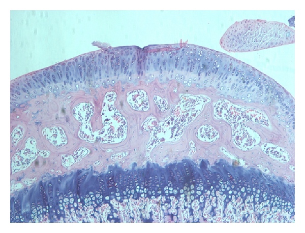
About histological analysis, in the treated group, an average area of the cartilage damage was 62.60%, and there was 35.5% of neocartilage formation after 45 days ( ). While in the control group, average area of cartilage lesions was 53.13%, but only 15.5% of neocartilage formation after 45 days (Figure 3).

Figure 3: HE staining of articular cartilage. Mature cartilage layer was recorded in normal mouse (a). Mature cartilage was thinned by needle (b). Injured cartilage was regenerated in negative control group (c, e) and treated group (d, f). However, the neocartilage in treated group was thicker than in negative control group.

The grade of cartilage injury between two experimental groups was different due to the effects of the dissimilar force from needle. After 45 days, results showed that 35.5% of neocartilage formed in treated group, while only 15.5% of neocartilage formed in the negative control group. This suggested that SVF and PRP gave benefit effects on the enhancement as well as trigger the neocartilage forming. More importantly, the articular cartilage in both of groups completed at the same level after 45 days with 12 cell layers. These results demonstrated that the SVF and PRP could participate in the process of self-renewal of joint cartilage at the joint microenvironment. Especially, there were no scar tissues or tumors forming at the graft sites. This result was similar to the previous publications about SVF plus PRP transplantation in the treatment of cartilage injury in dog [3133], rabbits [34, 61], horses [14, 61], rat [35], mice [36], and goats [37]. For example, in joint injured mice model by collagenase, Ter Huurne et al. (2011) showed that the level of damage nearly 50% reduction in ADSC transplanted mice compared to control after 42 days [36]. Specifically, knee injury went down to 25% in treated mice compare to 88% in controls. They suggested that the transplanted ADSC protected and healed of injured cartilage [37]. The findings of Dragoo et al. (2007) showed that autologous ADSCs could reestablish the joint surface in rabbits, in which 100% of rabbits (12/12) had the occurrence of neocartilage, while only 8% rabbit (1/12) in the control had the appearance of neocartilage ( ) [62].
Roles of SVF or ADSCs in cartilage regeneration were recorded with many different effects. In fact, similar to MSCs derived from bone marrow, ADSC had anti-inflammatory properties [63, 64] and inhibition of graft versus host disease (GVHD) [65]. The transplantation of ADSC could successfully treat graft versus host disease with steroid-resistant form [66, 67]. All of these roles could add more effects to trigger rapid cartilage regeneration in this study.

Besides, in this study, ingredients from PRP also had important roles in stimulated grafted cells as well as endogenous cells growth and differentiation. There are at least six known growth factors such as platelet-derived growth factor (PDGF) that promotes blood vessel growth, cell division, and forming the skin; transforming growth factor-beta (TGF-b) that promotes cell division mitosis and bone metabolism; vascular endothelial growth factor (VEGF) that promotes the blood vessel formation; epidermal growth factor (EGF) that promotes cell growth and differentiation, angiogenesis, and collagen formation; fibroblast growth factor-2 (FGF-2) that promotes the growth of cell differentiation and angiogenesis; and insulin-like growth factor (IGF) that is a regulator in all cell types of the body [68, 69]. PRP injection also showed that improvements in knee injury and osteoarthritis score, including pain and symptom relief [70, 71].

Combining effect of SVF and PRP has a positive effect on the stimulation of proliferation, differentiation, and regeneration of cartilage in a mouse model. However, SVF also has a few limitations, notably the relatively low presence of ADSCs in the SVF. Therefore, SVF cultured to enrich ADSC before being transplanted may be essential, especially a little obtained fat cases.

4. Conclusion

Adipose tissue provides a rich source of MSCs. The SVF and PRP injection are a promising therapy in injured articular cartilage regeneration. This therapy significantly improved the injured articular cartilage. However, this study only assesses the ability of tumorigenicity and efficiency in mouse. Some side effects such as fever and muscle pain as well as the tumorigenicity in human being when using SVF and PRP could not be checked in this research.

Abbreviations

SVF :

Stromal vascular fraction

PRP :

Platelet-rich plasma

RT-PCR :

Reverse transcription polymerase chain reaction

NOD/SCID :

Nonobese diabetic/severe combined immunodeficient

SSEA :

Stage-specific embryonic antigen

MSC :

Mesenchymal stem cell

ADSC :

Adipose-derived stem cell

D-PBS :

Dulbecco-modified phosphate buffered saline

HE :

Hematoxylin and eosin

ESC :

Embryonic stem cell

GVHD :

Graft versus host disease.


Charlie 發表在 痞客邦 留言(0) 人氣()

832396.fig.001a832396.fig.002a  

Transplantation of Non-expanded Adipose Stromal Vascular Fraction and Platelet-Rich Plasma for Articular Cartilage Injury Treatment in Mice Model1

Journal of Medical Engineering
Volume 2013 (2013), Article ID 832396, 7 pages
http://dx.doi.org/10.1155/2013/832396

Research Article

Phuc Van Pham,1 Khanh Hong-Thien Bui,2 Dat Quoc Ngo,3 Lam Tan Khuat,1 and Ngoc Kim Phan1

1Laboratory of Stem Cell Research and Application, University of Science, Vietnam National University, Ho Chi Minh City, Vietnam
2University of Medical Center, Ho Chi Minh University of Medicine and Pharmacy, Ho Chi Minh City, Vietnam
3Department of Pathology, University of Medicine and Pharmacy, Ho Chi Minh City, Vietnam

Received 13 August 2012; Accepted 25 December 2012

Academic Editor: Ayako Oyane

Copyright © 2013 Phuc Van Pham et al. This is an open access article distributed under the Creative Commons Attribution License, which permits unrestricted use, distribution, and reproduction in any medium, provided the original work is properly cited.

Abstract

Stromal vascular fraction (SVF) combined with Platelet-rich plasma (PRP) is commonly used in preclinical and clinical osteoarthritis as well as articular cartilage injury treatment. However, this therapy has not carefully evaluated the safety and the efficacy. This research aims to assess the safety and the efficacy of SVF combined with PRP transplantation. Ten samples of SVFs and PRPs from donors were used in this research. About safety, we evaluate the expression of some genes related to tumor formation such as Oct-4, Nanog, SSEA3, and SSEA4 by RT-PCR, flow cytometry, and tumor formation when injected in NOD/SCID mice. About efficacy, SVF was injected with PRP into murine joint that caused joint failure. The results showed that SVFs are negative with Oct-4, Nanog, SSEA-3, and SSEA-4, as well as they cannot cause tumors in mice. SVFs combined with PRP can improve the joint regeneration in mice. These results proved that SVFs combined with PRP transplantation is a promising therapy for articular cartilage injury treatment.

1. Introduction

Stem cell therapy is considered as a promising therapy for degenerative disease treatment, especially articular cartilage injury as well as osteoarthritis. Osteoarthritis was treated by stem cell transplantation for a few years ago. Stem cells from various sources were used to treat this disease. However, the mesenchymal stem cells (MSCs) are considered as most suitable candidates. MSCs are multipotential cells capable of differentiation into bone, cartilage, fat, and some other cells [1]. MSCs could be isolated from Bone marrow [2], Adipose tissue [3], Cord blood [4], Banked umbilical cord blood [5], Umbilical cord [6], Wharton’s jelly [7], Placenta [8], and Pulp [9]. However, MSCs from bone marrow [1012] and from adipose tissue [1315] are two common stem cell sources for treating cartilage degeneration.

Cartilage degeneration or cartilage injury is a common clinical problem and easily leads to osteoarthritis. Osteoarthritis is a chronic degenerative process characterized by the degeneration of cartilage, bone bud formation, cartilage reorganization, joint erosion, and loss of joint function [16]. Currently, cartilage injury was treated primarily with drugs [1720] or injection of hyaluronic acid [21, 22] to reduce the symptoms, pain, and inflammation control. However, these therapies’ efficiencies were limited and often failed to prevent the degeneration of the joints [23].

MSCs from adipose tissue, also known as stem cells isolated from fat tissue (Adipose-derived stem cellsADSCs), are a suitable source of Mesenchymal stem cells for Autograft. This stem cell source was used to treat many diseases such as liver fibrosis [24], sciatic nerve defects [25], systemic sclerosis [26], ischemia [27], skeletal muscle injury [28], passive chronic immune thrombocytopenia [29], and infarcted myocardium [30]. Recently, they have been extended to treat cartilage injuries as well as osteoarthritis such as dogs [3133], rabbits [34], horses [14], rat [35], mice [36], and goats [37]. These researches demonstrated that Neocartilage formed after ADSC transplantation. Some phase I and II clinical trials using ADSCs transplantation are performed to treat osteoarthritis and cartilage degeneration (NCT01300598, NCT01585857, NCT01399749). Pak (2011) showed that all ADSC grafted patients improved the cartilage regeneration [15].

Among all of ADSC transplantation cases, SVF is used as Non-cultured ADSC (Non-expanded ADSC). SVF transplantation has some advantages such as saving time (from isolation to transplant faster about 23 hours), being inexpensive, and reducing the risk of cell culture. Although many studies have demonstrated the benefits of SVF/ADSC transplantation in cartilage injury treatment, especially knee articular cartilage, so far a little comprehensive studies aim to evaluate the safety and efficiency of SVF transplantation for articular cartilage treatment. Therefore, this study aims to evaluate the safety and efficiency of SVF transplantation combined with PRP in the treatment of cartilage injury in the mouse model.

2. Materials and Methods

2.1. SVF and PRP Preparation

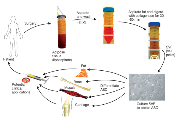
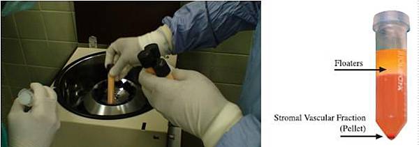
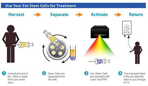
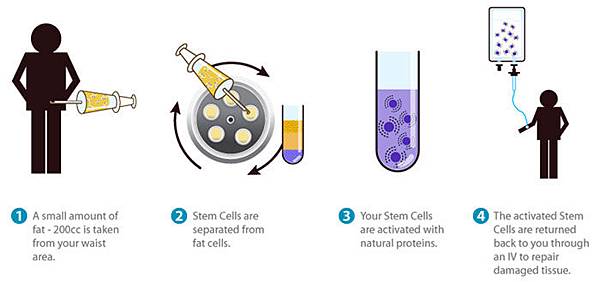
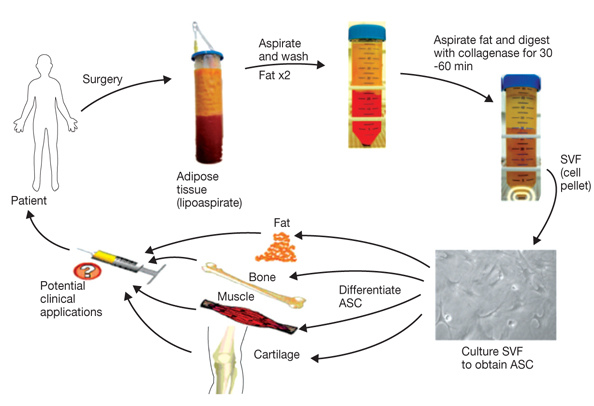
Firstly, adipose tissue was collected from abdominal fat tissue of ten consenting healthy donors. About 40–80mL ​​of fat was collected by syringe and stored in 100mL sterile bottle. Fat was kept at 2–8°C and then quickly moved to the laboratory. SVF cells are separated from the fat using the extraction kits (Adistem, Australia) according to the manufacturer’s guideline. Briefly, fat is Washed 3 times with saline solution to eliminate red blood cells. Then, the fat was Incubated with a solution AdiExtract (Adistem, Australia). The sample was Centrifuged to collect SVF as Pellet at the bottom of the tube. To prepare Platelet-rich plasma (PRP), 50mL of peripheral blood was taken from a large vein (arm veins). Blood was centrifuged 1,700rpm for 10 minutes to get platelet-enriching plasma. This plasma is Activated with Activator solution (Adistem, Australia). Then, PRP was Mixed with SVF to make the cell suspension. Finally, this suspension was Stimulated by the LED light (light monochromatic low energy, Adlight, Adistem, Australia) for 30 minutes before using for treatment.

2.2. Quantification of Nucleated Cells from SVF

Cell suspension (SVF and PRP) is used to count the nucleated cells. Cell number and percentage of viable cells were determined by automatically nucleus-based cell counter (NucleoCounter, Chemometec). Total cell numbers were counted after permeabilization of the membrane by Reagent A (lysis buffer, Chemometec) and neutralized with a solution of Reagent B (Neutralized buffer, Chemometec). Cell suspension is loaded into the counting chamber containing Propidium iodide dye. For counting dead cells, suspension cells were mixed with only Reagent B solution and loaded into the counting chamber. Survival rate is calculated as follows: (total cell number − the number of dead cells): the total number of cells × 100%.

2.3. Evaluation of the Existence of ADSC in SVF

The existence of ADSC in SVF determined by Flow cytometry. The process summarized as follows: cells were washed twice in physiological saline of Dulbecco-modified PBS (D-PBS) supplemented with 1% bovine serum albumin (Sigma-Aldrich, St Louis, MO). Cells were stained for 30min at 4°C with the monoclonal antibody anti-CD44-PE, anti-CD90-PE, and anti-CD105-FITC (BD Biosciences, Franklin Lakes, New Jersey offers). Stained cells were analyzed by flow cytometer FACSCalibur machine (BD Biosciences). Isotype control is used for all analyzes.

2.4. RT-PCR

To evaluate the safety of SVF, we should identify the gene expression levels related to the process of causing tumors and test the ability to form tumors in vivo. About gene expression, RNA was isolate by Trizol according to the manufacturer’s instructions (Sigma-Aldrich, St Louis, MO). RNA precipitated with isopropanol at room temperature for 10 minutes. ADSC cells analyzed the expression of genes related to markers of cancer cells or embryonic stem cells, Oct-3/4 and Nanog by Real-time kit SYBR RT-PCR one tube-one step (Sigma-Aldrich, St. Louis, MO). The used primers were Oct-3/4, forward primer: F: 5′-GGAGGAAGCTGACAACAATGAAA-3 ′, reverse primer R: 5′-GGCCTGCACGAGGGTTT-3; Nanog, forward primer: F: 5′-ACAACTGGCCGAAGAATAGCA-3′; reverse primer R: 5′-GGTTCCCAGTCGGGTTCAC-3; GAPDH, forward primer: F: 5′-GGGCTGCTTTTAACTCTGGT-3′; reverse primer: R: 5′-TGGCAGGTTTTTCTAGACGG-3′.

2.5. In Vivo Tumorigenicity Assay

The tumorigenicity of ADSC was evaluated in mice NOD/SCID (NOD.CB17-Prkdcscid/J, Charles River Laboratories). All mice manipulation was according to guideline of laboratory and approved by the Local Ethics Committee of Stem Cell Research and Application, University of Science (VNU-HCM, VN). All mice were kept in clean condition. Mice were injected subcutaneously at a concentration of 105, 106, and 107 cells, respectively in three groups (each group with 3 mice). Control group was injected with PBS. The formation of tumors in mice was followed for 3 months.

2.6. Articular Cartilage Injured Mice Model and Experimental Treatment Schedule

To evaluate the efficiency of SVF transplantation in articular cartilage injury, we used articular cartilage injured mouse model. The NOD mouse/SCID mice were anesthetized with ketamine (40mg/kg), then joint destruction by fine needle 32.5G. Normal mice were used as a positive control (uninjured). Nine mice randomly divided into the treatment group (5 mice) and negative control group (4 mice). Six hours after injury, the mice were treated. In the treatment group, 200μL containing SVF in PRP (the treatment group) or PBS (the negative control group) was injected into the knee joint via two doses, with a 10min interval between injections.

Mice were recorded some parameters related to joint regeneration for 45 days. The mice were recorded the movement on the table daily. At the 45th day, all mice were anesthetized, and their hind limbs were cut and used for histological analysis and further experiments. The samples were fixed in 10% formalin, decalcified, sectioned longitudinally, and stained with hematoxylin and eosin (HE) (Sigma-Aldrich, St Louis, MO). Using HE stained slides, three parameters were examined such as the area of injured cartilage (%), the area of neo-cartilage (%), and the number of neocartilage cell layers. The injured cartilage area was determined as the percentage of lost mature cartilage compared to the control. Data was analyzed using Statgraphics software (v7.0; Statgraphics Graphics System, Warrenton, VA).

3. Results and Discussion

MSCs have the large differentiative potential, easily differentiate into bone, cartilage, and adipocyte. Autologous MSC transplantation is considered as a safe and effective therapy in some patients. Recently, adipose tissue was identified as the abundant source of MSCs. ADSCs exist with large amounts of adipose tissue [38]. Similar to MSCs from other sources, ADSC have the ability to differentiate into fat cells, bone, and cartilage and transdifferentiate into neurons and muscle [3945]. Therefore, ADSC is favored as a source of autologous cell transplantation. However, the isolated ADSC relatively complex, consuming time, so ADSCs were mainly used as SVF (containing ADSC) without culture. This study aims to evaluate the safety and efficiency of SVF transplantation resuspended in PRP in mouse model.

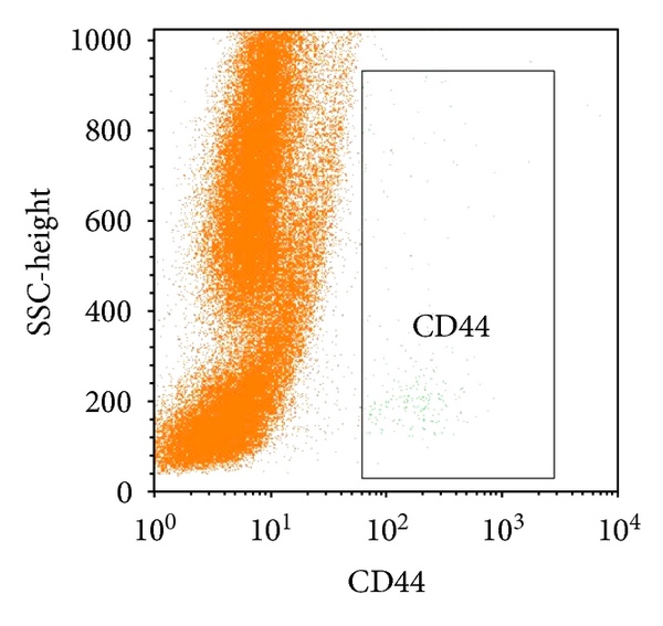
In the first experiment, we successfully isolated SVF and PRP. Compared to other studies, we have successfully isolated SVF cells from 1 gram of fat with a survival rate of % ( ). Next, we assessed the existence of ADSC or MSC in the SVF. Analysis results from 10 samples showed that ADSCs existed in all samples. ADSC counted to % in the SVF. ADSC populations were identified based on the expression of CD44, CD90, and CD105 of them (Figure 1). These results were similar to many other authors on ADSC markers [43, 4652]. The markers satisfied the criteria of MSC following to Dominici et al. (2006) [53].

Figure 1: Existence of ADSCs in SVF. ADSCs were confirmed based on expression of CD44, CD105 and CD90.

To assess safety, we have evaluated the expression of genes related to cancer. In particular, two genes Oct-3/4 and Nanog were assessed by real-time RT-PCR method and SSEA-3, and SSEA-1 was assessed by flow cytometry. The results showed expression of Oct-3/4, Nanog, SSEA-3, and SSEA-1 much lower than embryonic stem cells (Figure 2). These results demonstrated that the SVF hold low tumorigenicity. In fact, Nanog and Oct-3/4 participate in the process of self-renewal of embryonic stem cells [54, 55]. Moreover, these proteins related to the tumorigenicity process in mature germ cells [56], carcinoma oral squamous cell [57], lung cancer [58], breast cancer [59], and gliomas [60]. The SVF and PRP injection under the skin mouse NOD/SCID could not form teratomas. With these experiments, we concluded that the SVF plus PRP has a promising therapy with a high safety for transplantation experiments.

Figure 2: Expression of Oct-3/4, Nanog, SSEA-3, and SSEA-1 in SVF. Oct-3/4 and Nanog expressed lower in embryonic stem cell (ESC) (a); while SSEA-3 and SSEA-1 did not express in SVF (b and c).

Charlie 發表在 痞客邦 留言(0) 人氣()

Regeneration of human bones in hip Osteonecrosis and human cartilage in knee Osteoarthritis with autologous Adipose-tissue-derived stem cellsa case series5

Discussion

This series of clinical case reports provides clear MRI evidence of apparent bone regeneration in osteonecrosis of femoral heads and meniscus cartilage regeneration in osteoarthritis of human knees. Based on the MRI features, it is probable that the new tissue formation is bone matrix in the case of osteonecrosis and meniscus cartilage in osteoarthritis. However, without biopsy, the true nature of the newly-formed tissue is unclear. While bone and cartilage regeneration using ADSCs has been shown in animal models, these case reports represent the first successful regeneration of bones and cartilage in human patients.

In addition to the MRI evidence, the patients' symptoms and signs also improved. It is worthwhile to note that the patients' symptoms improved gradually over three months. Thus, it can be speculated that, in patients with osteonecrosis, newly-formed bone has concomitant neovascularization. Osteonecrosis, or avascular necrosis, occurs due to compromise in blood circulation. Without concurrent neovascularization, the consolidation or regeneration of bones cannot be sustained.

Another issue with these clinical results is that patients with osteoarthritis did not report 100% symptom improvements. This may be due to the fact that osteoarthritis is a disease of the whole knee, not just the cartilage.

With regard to the mechanism of tissue regeneration, there are a few plausible possibilities. The mechanism of regeneration could be through direct differentiation of stem cells that were introduced through the injection. However, there is a possibility that the ADSCs exert tropic effects on the existing tissues as well. Numerous studies have reported that MSCs, in addition to tissue repair and regenerative effects, have immunomodulatory and paracrine effects [14].

Furthermore, PRP could have contributed to the regeneration of bones and blood vessels. PRP contains multiple growth factors including TGFβ, IGF, FGF, and PDGF. A literature review of the data on the uses of PRP showed that it has a positive effect on the stimulation of bones and blood vessels and chondrocytes. Here, it was used as a growth factor and as a differentiating agent for the MSCs.

Further, Dexamethasone injection, used as a differentiating agent for Cartilage, may also have had positive effects in patients with osteoarthritis. The levels injected (100 ng/mL) were negligible compared to the doses being used in clinical settings. Such low doses in the nanogram range have been shown to increase extracellular matrix production by chondrocytes, and are commonly used in vitro to differentiate MSC from cartilage [15].

This is the first series of case reports showing possible successful bone and cartilage regeneration in humans by using a combination of ADSCs, hyaluronic acid, PRP and CaCl2. Currently, no non-surgical therapy is available for the treatment of osteonecrosis and osteoarthritis. Thus, stem cell therapy may significantly improve current treatment strategies for the treatment of knee osteoarthritis and osteonecrosis of the femoral head. However, further studies need to be initiated to find out the true detailed nature of the apparently regenerated bones and cartilage and to determine the true mechanism of tissue regeneration.


Conclusions

After three months of treatment, all the patients reported on above were able to straighten their hips and extend their knees further, affecting MRI postures. Therefore, obtaining the post-treatment MRI data at the exactly same location as pre-treatment MRI of the hips and knees was difficult.

Although there were difficulties in repeatedly obtaining the exact location of the hips and knees, the pre-procedure and post-procedure MRI analyses clearly demonstrate filled bone defects in osteonecrosis and increased meniscus cartilage volume in osteoarthritis, indicating regeneration attributable to the ADSC treatment. Additionally, the measured physical therapy outcomes, subjective pain, and functional status, all improved


Consent

Written informed consent was obtained from all patients for publication of this case report and any accompanying images. Copies of the written consents are available for review by the Editor-in-Chief of this journal.


Competing interests

The authors declare that they have no competing interests.


Authors' contributions

JP was in charge of patient treatment and follow-up, was responsible for manuscript drafting and revision, and read and approved the final manuscript.


Acknowledgements

JP acknowledges the support from the staff of Miplant Stems Clinic.


References

Korean Food and Drug AdministrationCell therapy. In Korean Food and Drug Administration Rules and Regulations. Seoul, KoreaFood and Drug Administration; Chapter 2, Section 12

Arnoczky SP: Building a meniscus. Biologic considerations.

Clin Orthop Relat Res 1999, 367(Suppl):S244-S253. PubMed Abstract

Barry FP: Mesenchymal stem cell therapy in joint disease.

Novartis Found Symp 2003, 249:86-96. PubMed Abstract

Zhang HN, Li L, Leng P, Wang YZ, Lv CY: Uninduced adipose-derived stem cells repair the defect of full-thickness hyaline cartilage.

Chin J Traumatol 2009, 12:92-97. PubMed Abstract

Centeno CJ, Buse D, Kisiday J, Keohan C, Freeman M, Karli D: Increased knee cartilage volume in degenerative joint disease using percutaneously implanted, autologous mesenchymal stem cells.

Pain Physician 2008, 11:343-353. PubMed Abstract | Publisher Full Text

Lee JS, Lee JS, Roh HL, Kim CH, Jung JS, Suh KT: Alterations in the differentiation ability of mesenchymal stem cells in patients with nontraumatic osteonecrosis of the femoral head: comparative analysis according to the risk factor.

J Orthor Res 2006, 24:604-609. Publisher Full Text

Nuttelman CR, Tripodi MC, Anseth KS: In vitro osteogenic differentiation of human mesenchymal stem cells photoencapsulated in PEG hydrogels.

J Biomed Mater Res A 2004, 68:773-782. PubMed Abstract | Publisher Full Text

Parsons P, Hesselden K, Butcher A, Maughan J, Milner R, Horner A: The biological effect of platelet rich-plasma on the fracture healing process.

J Bone Joint Surg Br 2009, 91B(Suppl 2):293-c.

Li N, Yuan R, Chen T, Chen L, Jin X: Effect of platelet-rich plasma and latissimus dorsi muscle flap on osteogenesis and vascularization of tissue-engineered bone in dog.

J Oral Maxillofac Surg 2009, 67:1850-1858. PubMed Abstract | Publisher Full Text

Wu W, Chen F, Liu Y, Ma Q, Mao T: Autologous injectable tissue-engineered cartilage by using platelet-rich plasma: experimental study in a rabbit model.


J Oral Maxillofac Surg 2007, 65:1951-1957. PubMed Abstract | Publisher Full Text

Martineau I, Lacoste E, Gagnon G: Effects of calcium and thrombin on growth factor release from platelet concentrates: kinetics and regulation of endothelial cell proliferation.

Biomaterials 2004, 25:4489-4502. PubMed Abstract | Publisher Full Text

Zuk PA, Zhu M, Ashjian P, De Ugarte DA, Huang JI, Mizuno H, Alfonso ZC, Fraser JK, Benhaim P, Hedrick MH: Human adipose tissue is a source of multipotent stem cells.

Mol Biol Cell 2002, 13:4279-4295. PubMed Abstract | Publisher Full Text | PubMed Central Full Text

Peterkofsky B: Bacterial collagenase.

Methods Enzymol 1982, 82:453-471.

Salem H, Thiemermann C: Mesenchymal stromal cells: current understanding and clinical status.

Stem Cells 2010, 28:585-596. PubMed Abstract | Publisher Full Text | PubMed Central Full Text

Richardson DW, Dodge GR: Dose dependant effects of corticosteroids on the expression of matrix-related genes in normal and cytokine-treated articular chondrocytes.

Inflamm Res 2003, 52:39-49. PubMed Abstract | Publisher Full Text

Childs JD, Piva SR: Psychometric properties of the functional rating index in patients with low back pain.

Eur Spine J 2005, 14:1008-1012. PubMed Abstract | Publisher Full Text

Charlie 發表在 痞客邦 留言(0) 人氣()

1752-1947-5-296-51752-1947-5-296-6  

Regeneration of human bones in hip Osteonecrosis and human cartilage in knee Osteoarthritis with autologous Adipose-tissue-derived stem cellsa case series4

The final case concerns a 79-year-old Korean woman with over seven years' history of bilateral knee pain due to osteoarthritis.

Her left knee was much more painful than the right. Due to her occupation, she made active use of her bilateral knee joints. With a diagnosis of osteoarthritis of both knees, she had received multiple injections of steroids and hyaluronic acid in both knees over the years. However, she noticed no improvement of pain. She was seen by an orthopedic surgeon and was offered a TKR. She was also reluctant to go through the TKR due to possible side effects. Since then, she had been receiving physical therapy with little improvement.

At the time of initial evaluation, she reported severe pain in the left knee (VAS score 8) on rest. The pain was increased when walking. On physical examination, there was deformity of the knee, mild joint swelling, a decreased range of motion and tenderness with flexion. Apley and McMurray tests were negative, and there was no ligamentous laxity.

A pre-treatment 1.5T MRI demonstrated a decreased size and deformed contour on her medial meniscus of the left knee due to maceration. She also underwent the same procedure as our previous patient.

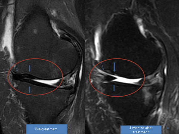
After the fourth week of ADSC injection, her pain improved over 50% and flexion of the knee improved as well. By week 12, her pain had improved over 90% and she was able to flex her knee further (Tables 7 and 8). A repeat MRI taken at week 12 showed a significant increase in the height of her meniscus cartilage on the anterior medial side of the left knee (Figures 5 and 6).


Table 7. Functional rating index and visual analog scale (VAS) score of patient 4

Table 8. Physical therapy (PT) range of motion of patient 4

Figure 5. MRI sagittal T2 view of the knee. Pre-treatment and post-treatment MRI shows increased height of medial meniscus cartilage. The articular cartilage also has a clearer marking, representing probable cartilage regeneration.

Figure 6. MRI coronal T2 view of the knee. The anterior medial meniscus has increased in height.

Charlie 發表在 痞客邦 留言(0) 人氣()

1752-1947-5-296-31752-1947-5-296-4  

Regeneration of human bones in hip Osteonecrosis and human cartilage in knee Osteoarthritis with autologous Adipose-tissue-derived stem cellsa case series3

The third case concerns a 70-year-old Korean woman with over five years' history of right knee pain due to osteoarthritis.

Due to her occupation, she made active use of her bilateral knee joints. With a diagnosis of osteoarthritis of the right knee, she had received multiple injections of steroids and hyaluronic acid over the years. However, she did not notice any improvement in her pain. She was seen by an orthopedic surgeon and was offered a total knee replacement (TKR). She was reluctant to go through the TKR procedure due to possible side effects. Since then, she has been receiving physical therapy with little improvement.

At the time of initial evaluation, she reported moderately severe pain (VAS score 7) on rest. Her knee pain increased when walking. She also complained of mild knee swelling. On physical examination, there was mild joint swelling, a decreased range of motion and tenderness with flexion. Apley and McMurray tests were negative, and there was no ligament laxity.

A pre-treatment 1.5T MRI scan demonstrated a decreased size and deformed contour on the medial meniscus of the left knee due to maceration.

After obtaining ADSCs and preparing PRP as described above for the first two patients, she was prepared for injection of the mixture into the joint.

In order to inject the stem cell and PRP mixture, she was first placed in a supine position with her right knee bent at 90°. After cleaning with povodine-iodine and draping with sterile drapes, her knee was anesthetized with 2% Lidocaine at the medial and lateral sides of the inferior patella. Using a 22-gauge 1-inch needle, 8.5 cm3 of ADSCs, PRP, Dexamethasone and Hyaluronic acid mixture was injected into the medial and the lateral sides of the knee.

She was then instructed to remain still for 30 minutes to allow for cell attachment. As she was subsequently discharged from the clinic, she was instructed to maintain activity as tolerable.

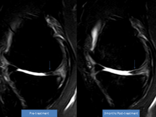
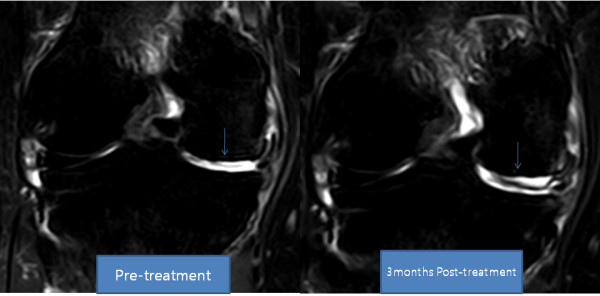
She returned to our clinic for Four additional PRP and Dexamethasone injections over the next four weeks. After the seventh week of ADSC injection, her pain had improved more than 80% and flexion of the knee had also improved. By week 12, her pain had improved more than 90% and the range of motion also further improved (Tables 5 and 6). A post-treatment MRI taken at week 12 showed a significant increase in the thickness of meniscus cartilage on the medial side of the right knee (Figures 3 and 4).


Table 5. Functional rating index and visual analog scale (VAS) score of patient 3

Table 6. Physical therapy (PT), range of motion of patient 3

Figure 3. MRI sagittal T2 view of the knee. Pre-treatment and post-treatment MRI shows increased height of medial meniscus cartilage and articular cartilage (arrow).

Figure 4. MRI coronal T2 view of the knee. Pre-treatment and post-treatment MRI shows increased height of medial meniscus (arrow).

Charlie 發表在 痞客邦 留言(0) 人氣()

1752-1947-5-296-2  

Regeneration of human bones in hip Osteonecrosis and human cartilage in knee Osteoarthritis with autologous Adipose-tissue-derived stem cellsa case series2

The second case concerns a 47-year-old Korean man who had been working as a diver until three years prior to presentation.

Approximately three years prior to presentation, he started having right hip pain and was diagnosed with osteonecrosis of the right hip. His pain had progressed over three years and he was offered a total hip replacement (THR). Being reluctant to proceed with the surgical procedure, he elected to proceed with stem cell treatment. Before the procedure, a repeat MRI of the hip was performed and a diagnosis of osteonecrosis of the femoral head, stage 4, was confirmed.

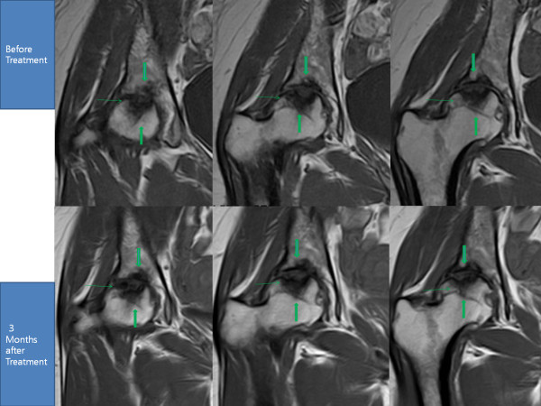
He then underwent the same procedures as our first patient. After the fourth week of the ADSC injection, his pain improved more than 30% along with improvement in range of motion. However, by week 12, his pain had only minimally improved further (Tables 3 and 4). Interestingly, a repeat MRI taken at week 12 showed a significant filling of bone defects with a possibility of bone matrix formation at the site of necrosis in the femoral head (Figure 2).


Table 3. Functional rating index and visual analog scale (VAS) score of patient 2

Table 4. Physical therapy (PT) range of motion of patient 2

Figure 2. MRI of the right hip; T1 sequential coronal views. The blue arrow shows the pattern of probable bone regeneration. The green arrow shows probable bone consolidation.

Charlie 發表在 痞客邦 留言(0) 人氣()

1752-1947-5-296-1  

Regeneration of human bones in hip Osteonecrosis and human cartilage in knee Osteoarthritis with autologous Adipose-tissue-derived stem cellsa case series1

Jaewoo Pak

CorrespondenceJaewoo Pak jaewoopak@paksmedical.com

Author Affiliations

Miplant Stems Clinic, 32-3 Chungdam-Dong, Gangnam-Gu, Fourth Floor, Seoul, Korea

Journal of Medical Case Reports 2011, 5:296

doi:10.1186/1752-1947-5-296


The electronic version of this article is the complete one and can be found online athttp://www.jmedicalcasereports.com/content/5/1/296

Received11 October 2010

Accepted7 July 2011

Published7 July 2011


© 2011 Pak; licensee BioMed Central Ltd.


This is an Open Access article distributed under the terms of the Creative Commons Attribution License (http://creativecommons.org/licenses/by/2.0), which permits unrestricted use, distribution, and reproduction in any medium, provided the original work is properly cited.


Abstract

Introduction

This is a series of clinical case reports demonstrating that a Combination of Percutaneously injected autologous Adipose-tissue-derived stem cells, Hyaluronic acid, Platelet rich plasma and Calcium chloride may be able to regenerate Bones in human Osteonecrosis, and with addition of a very low dose of Dexamethasone, Cartilage in human knee Osteoarthritis.


Case reports

Stem cells were obtained from adipose tissue of abdominal origin by digesting lipoaspirate tissue with collagenase. These Stem cells, along with Hyaluronic acid, Platelet rich plasma and Calcium chloride, were injected into the right hip of a 29-year-old Korean woman and a 47-year-old Korean man. They both had a history of right hip osteonecrosis of the femoral head. For cartilage regeneration, a 70-year-old Korean woman and a 79-year-old Korean woman, both with a long history of knee pain due to osteoarthritis, were injected with Stem cells along with Hyaluronic acid, Platelet rich plasma, Calcium chloride and a nanogram dose of Dexamethasone. Pre-treatment and post-treatment MRI scans, physical therapy, and pain score data were then analyzed.


Conclusions

The MRI data for all the patients in this series showed significant positive changes. Probable bone formation was clear in the patients with osteonecrosis, and cartilage regeneration in the patients with osteoarthritis. Along with MRI evidence, the measured physical therapy outcomes, subjective pain, and functional status all improved. Autologous Mesenchymal stem cell injection, in conjunction with Hyaluronic acid, Platelet rich plasma and Calcium chloride, is a promising minimally invasive therapy for Osteonecrosis of femoral head and, with Low-dose Dexamethasone, for Osteoarthritis of human knees.


Background

Adipose-tissue-derived stem cells (ADSCs) have been widely used in Korea over the last few years by plastic surgeons as a semi-permanent volume expander. In June 2009, the Korean Food and Drug Administration (KFDA) allowed ADSCs to be used as autologous cell transplant when obtained and processed within a medical clinic with minimal processing [1].

Mesenchymal stem cells (MSCs) are found in numerous human tissues including bone marrow, synovial tissue and adipose tissue. These have been shown to differentiate into bones, cartilage, muscle and adipose tissue, representing a promising new area of therapy in regenerative medicine [2].

Because of their potent capabilities, MSCs have been used successfully in animal models to regenerate cartilage and bones [3,4]. In 2008, Centeno and colleagues reported regeneration of knee cartilage in a human by using autologous culture-expanded bone-marrow-derived stem cells [5]. However, to the best of our knowledge ADSCs have never been used successfully in osteonecrosis of a femoral head and in osteoarthritis of a human knee.

Osteonecrosis, or avascular necrosis, of femoral head is relatively a common disorder affecting individuals in their 30s to 50s. Osteoarthritis of a knee is an even more common disorder, especially in older patients. Currently, the only cure for both diseases is surgical intervention. However, the successful regeneration of bones and cartilage with ADSCs may represent a promising new, minimally invasive, non-surgical alternative.

Many issues need to be resolved and clarified before the general application of the procedure. The mechanism of regeneration is not yet clear. It could be through direct differentiation of stem cells that were introduced to the diseased joints. Alternatively, it could be due to the tropic effects of ADSCs on the existing tissues. Further, various elements of the local environment can affect the differentiation of MSCs [6]. Also, it is believed that a scaffolding material might be needed to allow the MSCs to attach and engraft [7].

Platelet-rich plasma (PRP) was used as a growth factor and as a differentiating agent for the MSCs. PRP contains multiple growth factors including transforming growth factor(TGF)β, insulin-like growth factor (IGF), fibroblast growth factor (FGF), and platelet-derived growth factor (PDGF). A literature review of the data on PRP shows that it has a positive effect on the stimulation of Bones, Blood vessel and Chondrocyte formation [8-10]. Hyaluronic acid was added as a Scaffolding material, and Calcium chloride was used as a PRP-activating agent [11].

This series of case reports demonstrates successful clinical results of regenerating bones in osteonecrosis and cartilage in patients with osteoarthritis, using percutaneously implanted, autologous MSCs along with PRP, hyaluronic acid, calcium chloride (CaCl2) and very-low-dose dexamethasone.


Case presentations

The following cases concern four different individuals. Of the four, the first two cases involve bone regeneration in osteonecrosis of hips, the latter two cases regeneration of cartilage in osteoarthritis of knees.

The first case concerns a 29-year-old Korean woman with over a year's history of right hip pain due to osteonecrosis.

Approximately a year prior to presentation, she started having hip pain with no history of trauma. She was seen by a physician and was diagnosed with osteoarthritis of the hip after an MRI scan. After taking non-steroidal anti-inflammatory drugs (NSAIDs) for a few weeks, her hip pain improved. About a month prior to presentation, she again started having hip pain radiating to the anterior region of the right knee. The pain was worse when standing up, walking, and exercising. The pain improved with rest. However, this time, the pain was not greatly relieved with NSAIDs.

A repeat MRI showed osteonecrosis of the femoral head, stage 4. Since there is no effective non-surgical treatment of the disease, she elected to receive stem cell treatment.

At the time of initial evaluation, she reported moderately severe pain (visual analog scale (VAS) score 7) on rest, increased pain when standing and walking (VAS score 9).

For a week prior to liposuction, she was Restricted from taking corticosteroids, aspirin, NSAIDs, and oriental herb medications.

For the liposuction procedure, she was brought into an operating room and placed in a supine position. She was then sedated with Propofol 2 mg intravenously (push) and a 20 mg/hour rate of continuous infusion.

After cleaning her abdominal area with povodine-iodine and placing sterile drapes, an incision of approximately 0.5 cm was made approximately 5 cm below the umbilicus. Then, using Tumescent solution (500 cm3 Normal saline, 40 cm3 2% Lidocaine, 20 cm3 0.5% Marcaine, 0.5 cm3 Epinephrine 11000), the Lower abdomen area was anesthetized. Next, using a 3.0 Hartman cannula, a total of 160 cm3 of lipoaspirates were extracted and separated by gravity. The resulting 100 cm3 of adipose tissue was then Centrifuged at 3500 rpm for Five minutes. The end result was approximately 40 cm3 of packed adipose tissue, fibrous tissue, red blood cells and a small number of nucleated cells.

An equal volume of digestive enzyme, 0.07% collagenase type 1, composed of several collagenases, sulfhydryl protease, clostripain, a trypsin-like enzyme, and an amino peptidase, derived from Clostridium histolyticum (Adilase; Worthington, Lakewood, NJ, USA) was then mixed with the centrifuged lipoaspirates at a ratio of 11 and Digested for 30 minutes at 37°C while rotating [12].

Bacterial collagenases differ from vertebrate collagenases in that they exhibit broader substrate specificity [13].

After the digestion, the lipoaspirates were Centrifuged at 100g for three minutes to separate the lipoaspirate and the enzyme. The leftover enzyme was then removed.

Using 500 cm3 5% dextrose in lactated Ringer's solution, the lipoaspirates were washed three times to remove the collagenase. After each washing, the lipoaspirates were centrifuged at 100 g. After the last centrifuge process, approximately 10 cm3 of ADSCs were obtained.

While preparing the ADSCs, 30 cm3 of autologous blood was drawn with 2.5 cm3 of anticoagulant citrate dextrose solution (ACD) formula. This was Centrifuged at 200 g for Five minutes. The resultant supernatant was drawn and Centrifuged at 1000 g for Five minutes. The supernatant was drawn and discarded. The resulting buffy coat was mixed with 10 cm3 of ADSCs.

Hyaluronic acid 1 cm3 was added to this mixture to act as a scaffold. This PRP was again mixed with CaCl2 for activation of platelets at a ratio of 102 (PRP 102 CaCl2).

In order to inject the mixture of stem cells and PRP, our patient was first placed in a lateral position with her left side down. After cleaning with povodine-iodine and draping with sterile drapes, 2% Lidocaine was used to anesthetize the hip at the femoral head region. Using a 22-gauge 3.5-inch needle, 17 cm3 mixture of ADSCs, PRP, Hyaluronic acid and CaCl2 were injected into the femoral head under ultrasound guidance.

She was then instructed to remain still with her leg elevated for 30 minutes to allow for cell attachment. On discharge home, she was instructed to maintain activity as tolerated. She returned to the clinic for four additional PRP injections with Calcium chloride every week over a period of a month.

After the fourth week of the ADSC injection, her pain had improved more than 50%. By week 12, her pain had improved more than 70% along with an improvement in range of motion (Tables 1 and 2). A repeat MRI taken at week 12 showed a significant filling of bone defects on the superior acetabulum and probable bone matrix formation in the subcortical region of the femoral head (Figure 1).


Table 1. Functional rating index [16] and visual analog scale (VAS) score for patient 1

Table 2. Physical therapy (PT) range of motion of patient 1


Figure 1. MRI of the right hip; T1 sequential coronal views. The cavity surrounded by the three green arrows has decreased in size in post-treatment MRIs due to probable bone regeneration.

Charlie 發表在 痞客邦 留言(0) 人氣()

SVF  

Breaking down fatComposition of Stromal vascular fraction

by Alexey Bersenev

on May 18, 2013

http://stemcellassays.com/2013/05/breaking-fat-svf/


In the previous post we defined adipose-derived stromal vascular fraction (SVF) versus stromal (stem) cells and highlighted difference between them. Today we will look at SVF more precisely and define its composition.

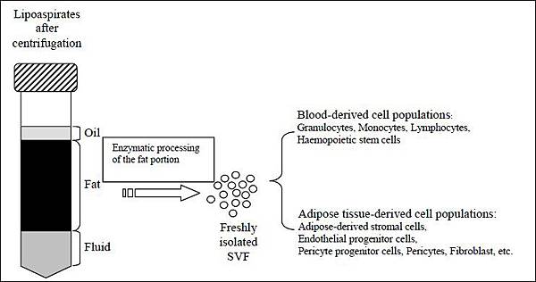
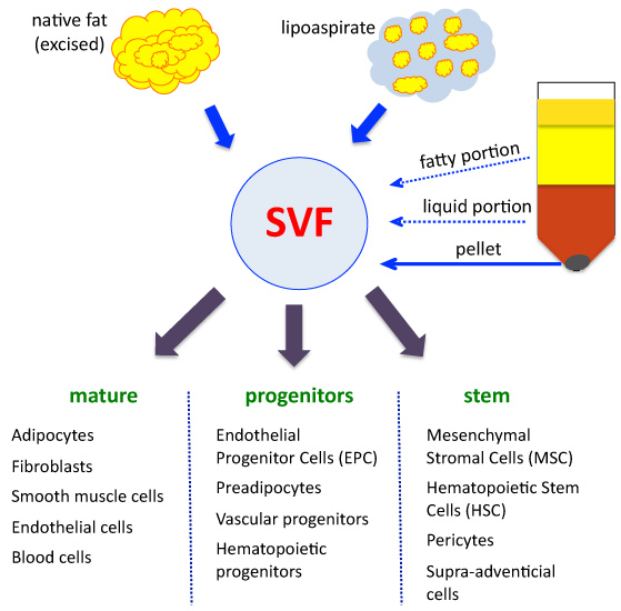
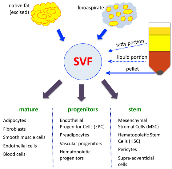
SVF is freshly isolated heterogeneous cell fraction, which could be derived from Native adipose tissue or Liposuction aspirates. SVF could be derived from both – the Fatty and Fluid portions of liposuction aspirates after enzymatic digestion. If SVF derived from fatty portion, it’s also called PLAprocessed lipoaspiratescells.

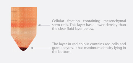
Basically, SVF is what remained in the pellet after removal blood and fat components. It is very crude and heterogeneous mix of multiple cell populations with different degree of maturity and function.

I draw a scheme of SVF isolation and composition

Based on method of adipose tissue processing, cellular composition of SVF can vary significantly. Most sources indicate that Adipose-derived stromalstemcells represent up to 10% (210%) of SVF. Endothelial cellsmature and progenitorscould represents anything from 7% up to 30% of SVF. Depending on processing, Fibroblasts could represent up to 50% of SVF (Cytori data, presented at ISCT 2010). CD34+ cells are present at large number and could compose up to 63% of SVF.

It has also been described that the SVF is composed of 11% CD2+ cells, 18% CD11a+ cells, 29% CD14+ cells, 49% CD31+ cells, 57% CD45+ cells, and 60% CD90+ cellsreferring to ASCs and endothelial cells. Others detected a different composition of the SVFnearly 11% CD14+ cells, ~2% CD31+ cells, ~7% CD34+, ~9% CD45+ cells, ~29% CD90+, and ~47% 146+ cells.

The SVF of human adipose tissue containedEndothelial progenitors, (15.4 ± 4.8)% (mean ± standard error), Pericytes (2.0 ± 1.1)%, CD146+/CD34+ transitional cells 0.5 ± 0.3, and SA-ASC (59.0 ± 10.0)% of Non-hematopoietic CD45−/CD14−/CD33−/glycophorin A−singlet cells.

To summarize

SVF composed of many Mature, Progenitor and Stem cell types. Depending on adipose tissue processing method, the composition of SVF and relative values of each cell population can vary significantly. Adipose-derived stromalstemcells represent 210% of SVF.

Charlie 發表在 痞客邦 留言(0) 人氣()

F1.largeScreen Shot 2012-03-19 at 1.20.30 PMSourcesofStemCells_2colb

StemCellRiskstem-cell-therapy  

Charlie 發表在 痞客邦 留言(0) 人氣()

01_adipose_diagram_02300441_1_En_146_Figa_HTMLadipose-derived-adult-stem-cellshighconcentrationijps_2012_45_2_229_101285_f2jkma-55-757-g001-lnri2921-f2separate_fatSVF螢幕擷取畫面 (63)  

Charlie 發表在 痞客邦 留言(0) 人氣()

AMS-2-8-g001AMS-2-8-g003fig-5螢幕擷取畫面 (59)螢幕擷取畫面 (60)螢幕擷取畫面 (61)螢幕擷取畫面 (62)  

Charlie 發表在 痞客邦 留言(0) 人氣()

000bottom_procedureDB_Pro_StemCell_HowDoStemCellsWorke_img_ourservice_01jkma-55-757-g001-lSVFsvns螢幕擷取畫面 (52)螢幕擷取畫面 (57)螢幕擷取畫面 (58)  

Charlie 發表在 痞客邦 留言(0) 人氣()

400px-Missingself.svgF1.largeF4.largefimmu-02-00088-g003fimmu-02-00088-g004fimmu-04-00372-g001ni.2032-F2螢幕擷取畫面 (55)螢幕擷取畫面 (56)  

Charlie 發表在 痞客邦 留言(0) 人氣()

284BB557-1D4F-4683-A31B-2F93BF9F62EE800px-Cancer_stem_cells_text_resized.svg23050414105ae.2Cancer Stem Cell Therapy(1)figure9_1illustration eurostem cell 02 therapy.img_assist_custom-479x338nrc1232-f2  

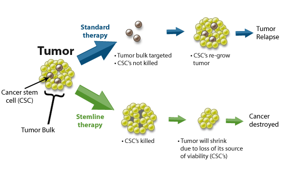
Cancer stem cellsCSCs癌症幹細胞and Stemline therapy or CSC-targeted cancer therapy

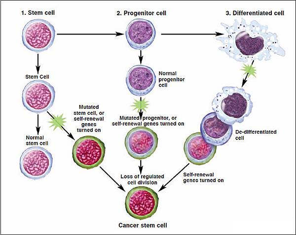
Cancer stem cellsCSCsare cancer cells found within tumors or hematological cancers that possess characteristics associated with normal stem cells, specifically the ability to give rise to all cell types found in a particular cancer sample.

CSCs are therefore tumorigenictumor-forming, perhaps in contrast to other non-tumorigenic cancer cells.

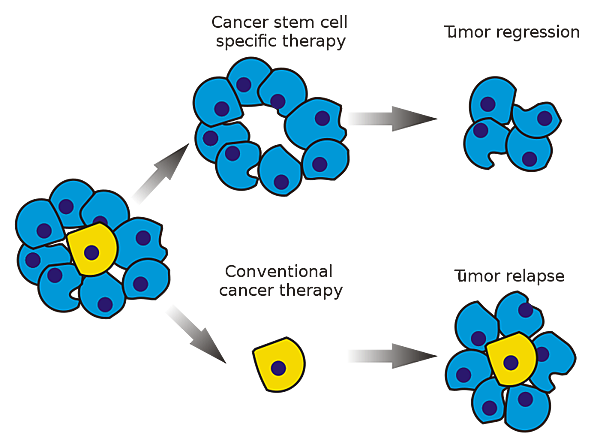
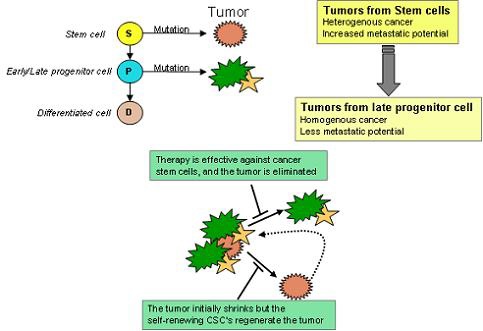
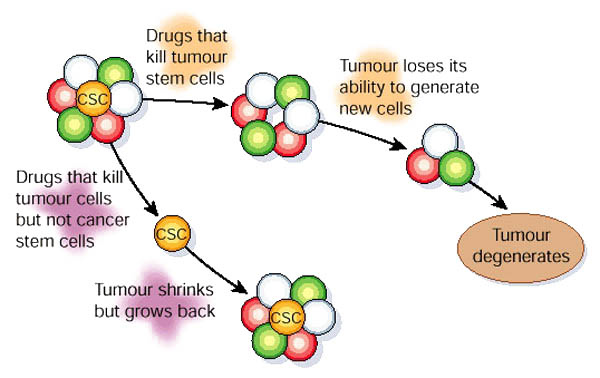
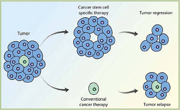
CSCs may generate tumors through the stem cell processes of self-renewal and differentiation into multiple cell types. Such cells are proposed to persist in tumors as a distinct population and cause relapse and metastasis by giving rise to new tumors. Therefore, development of specific therapies targeted at CSCs holds hope for improvement of survival and quality of life of cancer patients, especially for sufferers of metastatic disease.

Existing cancer treatments have mostly been developed based on animal models, where therapies able to promote tumor shrinkage were deemed effective. However, animals could not provide a complete model of human disease. In particular, in mice, whose life spans do not exceed two years, tumor relapse is exceptionally difficult to study.

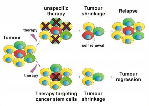
The efficacy of cancer treatments is, in the initial stages of testing, often measured by the ablation fraction of tumor massfractional kill. As CSCs would form a very small proportion of the tumor, this may not necessarily select for drugs that act specifically on the stem cells. The theory suggests that Conventional chemotherapies kill Differentiated or Differentiating cells, which form the bulk of the tumor but are unable to generate new cells. A population of CSCs, which gave rise to it, could remain untouched and cause a relapse of the disease.

癌症幹細胞(Cancer Stem CellCSC),又稱癌幹細胞、腫瘤幹細胞,是指具有幹細胞(Stem cell性質的癌細胞,也就是具有「自我複製」(self-renewal以及「具有多細胞分化」(differentiation等能力。通常這類的細胞被認為有形成腫瘤,發展成癌症的潛力,特別是隨著癌症轉移出去後,產生新型癌症的來源。

腫瘤內許多的細胞,其實只有一群細胞具有永生不死、持續分裂、分化的能力,而這些細胞,就稱為癌症幹細胞。

癌症幹細胞被認為是造成癌症轉移、復發,或是腫瘤對於化療、放射性療法產生抗性的原因之一。目前針對這個理論,學者希望研究出針對癌症幹細胞的療法,能夠專一性地殺死癌症幹細胞,以降低腫瘤產生抗藥性或是轉移的現象。

目前的癌症療法都是基於動物實驗的結果,目前的認知是只要癌症療法能將動物身上的腫瘤有效地縮小,就會被認為有其療效,然而要將實驗結果對照到人體醫療上仍有相當大的差距。比如說,以小鼠為例,生活週期很難超過兩年,如此短的生活週期無法提供癌症復發的動物模型可供研究。

而癌症幹細胞的理論認為,因為現行的治療都無法專一性的針對癌症幹細胞,而癌症幹細胞在腫瘤裡其實只佔一小部分,只要化療、放射性療法沒有殺死所有的癌症幹細胞,就會有抗藥性或是復發的風險出現。

Charlie 發表在 痞客邦 留言(0) 人氣()

588101-fig220121105163636_10472

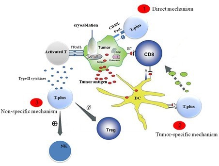
adoptF1.largeimage1image3immunotherapy-3nature10673-f2.2Ragde_Bahn_CryoFig4julbanner  

Charlie 發表在 痞客邦 留言(0) 人氣()

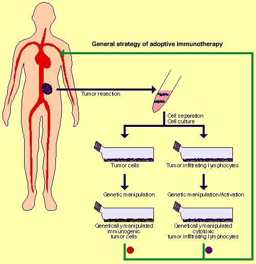
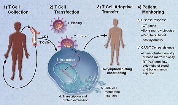
adoptAdoptive-cell-transfer1image1image2nrc1167-i1nrc2355-f1nrc478723.fig2WilsonWolf_PictureA_CellTherapyDiagram1812412172  

Adoptive cell transfer

Adoptive cell transfer can refer to either the transfer of cells, most commonly immune-derived cells, back into the same patient or into a new recipient host with the goal of transferring the immunologic functionality and characteristics into the new host.

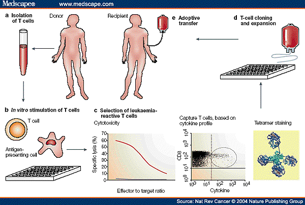
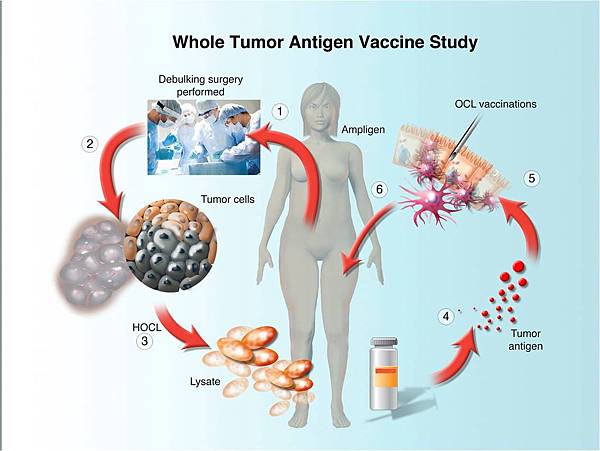
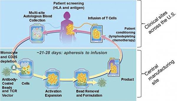
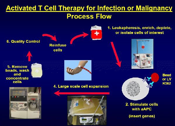
If possible, use of autologous cells helps the recipient by minimizing GVHD issues. For isolation of immune cells for adpotive transfer, a phlebotomist draws blood into tubes containing anticoagulant and the PBMbuffy coatcells are isolated, typically by density barrier centrifugation. In T cell-based therapies, these cells are expanded in vitro using cell culture methods relying heavily on the Immunomodulatory action of Interleukin-2 and returned to the patient in large numbers intravenously in an activated state. Anti-CD3 antibody is commonly used to promote the proliferation of T cells in culture. Research into interleukin-21 suggests it may also play an important role in enhancing the efficacy of T cell based therapies prepared in vitro.

An emerging treatment modality for various diseases is the transfer of stem cells to achieve therapeutic effect. Clinically, this approach has been exploited to transfer either Immune-promoting or Tolerogenic cellsoften lymphocytesto patients to either enhance immunity against viruses and cancer or to promote tolerance in the setting of autoimmune disease, such as Type I diabetes or rheumatoid arthritis.

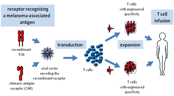
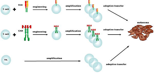
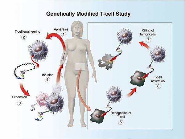
Cells used in adoptive therapy may be genetically modified using recombinant DNA technology to achieve any number of goals. One example of this in the case of T cell adoptive therapy is the addition of Chimeric antigen receptors, or CARs, to redirect the specificity of cytotoxic and helper T cells.

Charlie 發表在 痞客邦 留言(0) 人氣()

cancer_cell3-e1346685910571CAR-Engineered_T-Cell_Adoptive_Transferimage1image2nrc478723.fig2Picture3Picture4Picture6tcell_therapy-e1347441400780tech3WilsonWolf_PictureA_CellTherapyDiagram1812412172  

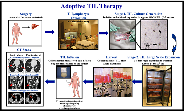
Adoptive T Cell Therapy

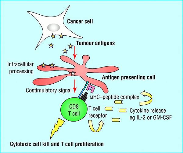
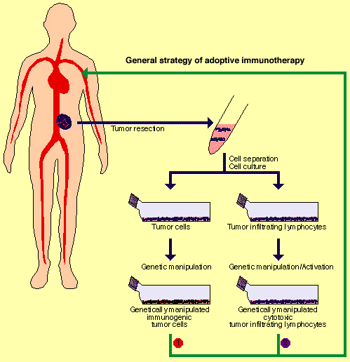
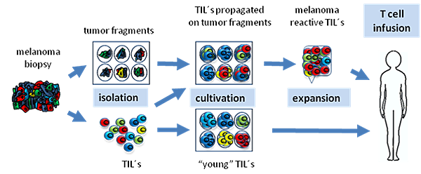
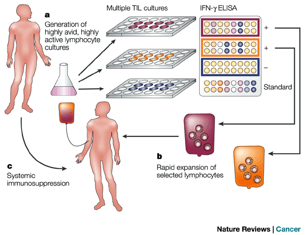
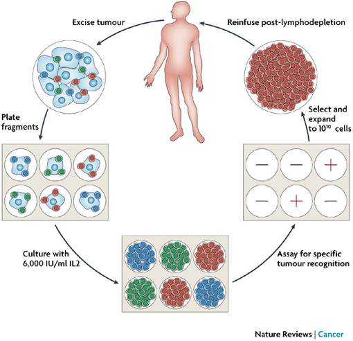
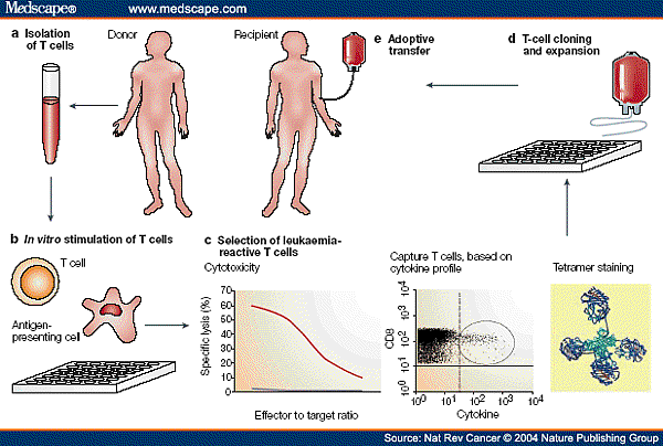
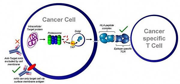
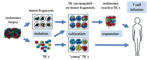
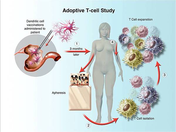
Adoptive T cell therapy involves the isolation and ex vivo expansion of tumor specific T cells to achieve greater number of T cells than what could be obtained by vaccination alone.

The tumor specific T cells are then infused into patients with cancer in an attempt to give their immune system the ability to overwhelm remaining tumor via T cells which can attack and kill cancer.

There are many forms of adoptive T cell therapy being used for cancer treatment; culturing tumor infiltrating lymphocytes or TIL, isolating and expanding one particular T cell or clone, and even using T cells that have been engineered to potently recognize and attack tumors.

One approach has been to utilize T cells taken directly from the patient’s blood after they have received a cancer vaccine.

A unique aspect to adoptive T cell therapy is the use of tumor specific CD4+ Th1 cells which may enhance anti-tumor efficacy.

Adoptive T cell therapy strategies have largely focused on the infusion of tumor antigen specific cytotoxic T cellsCTLwhich can directly kill tumor cells. However, CD4+ Th cells have a broader functionality. Th can activate antigen-specific effector cells and recruit cells of the innate immune system such as macrophages and dendritic cells to assist in antigen presentationAPC. Moreover, antigen primed Th cells can directly activate tumor antigen-specific CTL. In addition to direct contact, Th can activate CTL through cytokines such as IL-2 which stimulate the growth and expansion of effector T cells. In addition, Th1 induce the production of opsonizing antibodies that enhance the uptake of tumor cells into APC. These activated APC can then directly present tumor antigens to T cells.

As a direct result of activating APC, antigen specific Th1 have been implicated as the initiators of epitope or determinant spreading which is a broadening of immunity to other antigens in the tumor. The phenomenon of epitope spreading has been linked with a survival benefit after immunotherapy in patients with melanoma and breast cancer. The ability to elicit epitope spreading broadens the immune response to many potential antigens in the tumor and presumably would result in more efficient tumor cell kill due to the ability to mount a heterogeneic response. In this way, adoptive T cell therapy can used to stimulate endogenous immunity.

Charlie 發表在 痞客邦 留言(0) 人氣()

876203.fig.001BovenschenFigurewebsiteGranzyme_BGranzyme-B Pathwayni0203-106-F1nri911-f2nri911-f3nri1083-f3  

GranzymesSerine proteases顆粒溶解酶

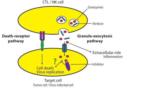
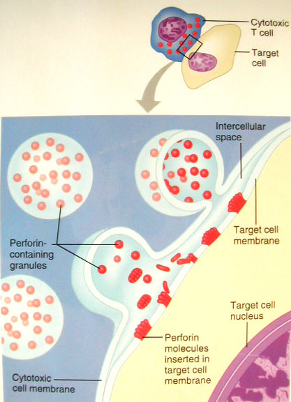
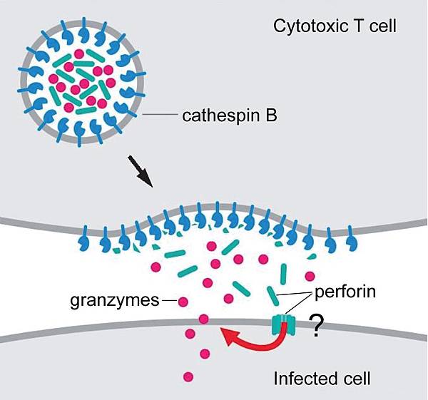
Granzymes are Serine proteases that are released by Cytoplasmic granules within Cytotoxic T cells and Natural killer cells.

Their purpose is to induce apoptosis within virus-infected cells, thus destroying them.

Granzymes when in the host cell are contained in cytotoxic granules to prevent harm to the host cell. Other locations that granzymes can be detected are in the rough endoplasmic reticulum, Golgi complex, and the trans-Golgi reticulum. The goal of the granules and perforins is to create a path way for the granzymes to follow and enter the target cells cytosol.

Granzymes are identified as being part of the serine esterase family.

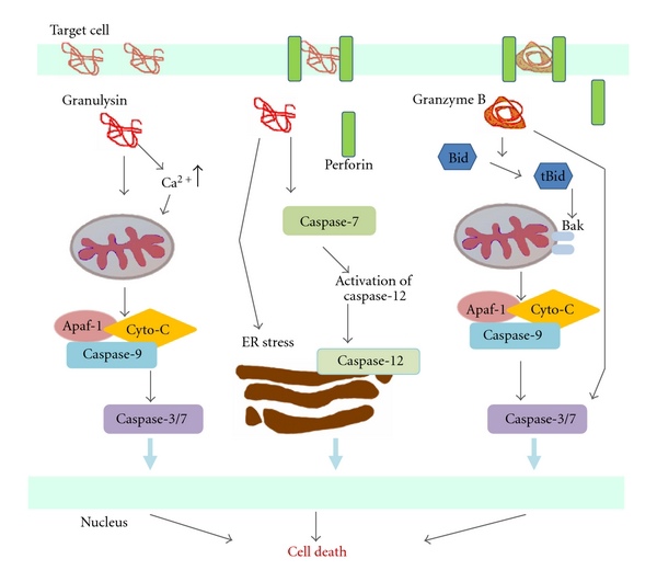
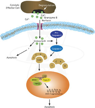
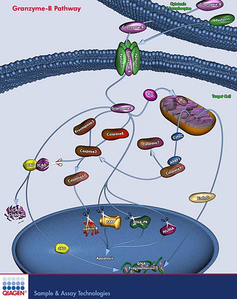
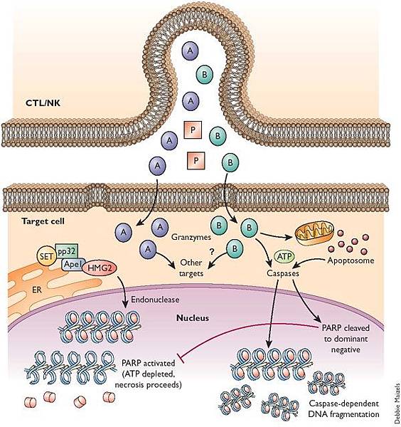
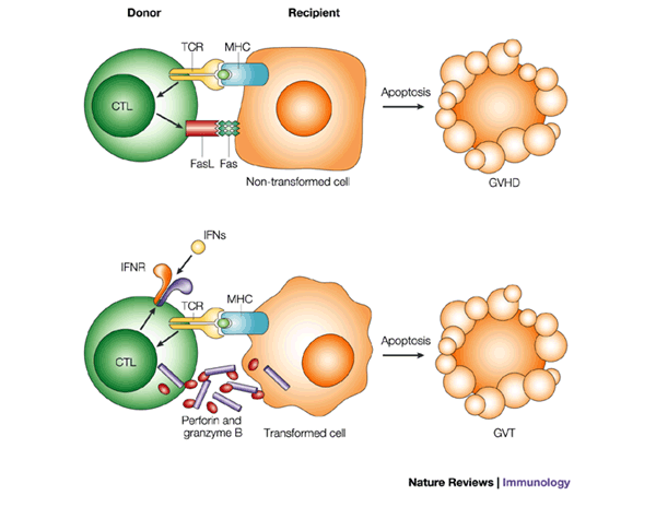
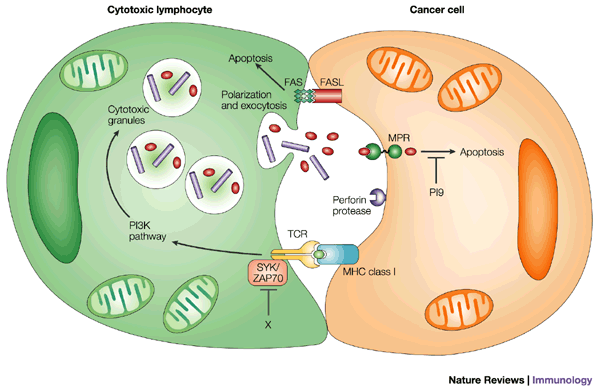
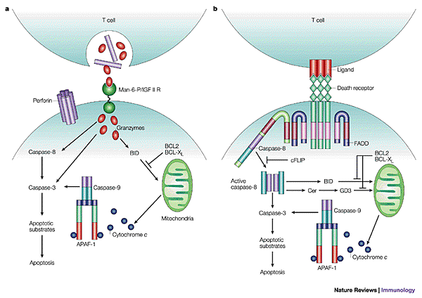
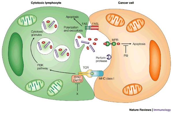
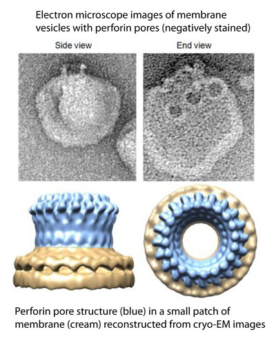
Cytotoxic T cells and Natural killer cells release a protein called Perforin, which attacks the target cells. Researchers used to think that perforin creates pores within the cell membranes, through which the granzymes can enter, inducing apoptosis. However, new evidence indicates that a multimeric complexGranzyme B, Perforin, and granulysincan enter a cell through the Mannose 6-phosphate receptor or another receptor found in tumor cells and is enclosed in a vesicle or a sac. Perforin then allows GrB to pass through the vesicle surface and into the cell, causing apoptosis by various pathways.

They do so by cleaving caspases, especially Caspase-3, which in turn activates Caspase-activated DNase. This enzyme degrades DNA, thus inducing apoptotic cascades. Also, GrB cleaves the protein Bid, which recruits the protein Bax and Bak to change the membrane permeability of the mitochondria, causing the release of cytochrome cwhich is one of the parts needed to activate caspase-9 via the Apoptosome, Smac/Diablo and Omi/HtrA2which suppress the Inhibitor of apoptosis proteinsIAPs, among other proteins. As well, GrB is shown to cleave many of the chemicals responsible for apoptosis without the aid of caspase, as proven by experiments on caspase knockout mice CTL cells incubated with other cells.

In 1986 Jürg Tschopp and his group published a paper on their discovery of granzymes. In the paper they discussed how they purified, characterized and discovered a variety of granzymes found within cytolytic granules that were carried by cytotoxic T lymphocytes and natural killer cells. Jürg was able to identify 8 different granzymes and discovered partial amino acid sequences for each. The molecules were unofficially named Grs for five years before Jürg and his team came up with the name granzymes which was widely accepted by the scientific community.

Granzyme secretion can be detected and measured using Western Blot or ELISA techniques. Granzyme secreting cells can be identified and quantified by flow cytometry or ELISPOT. Alternatively, granzyme activity can be assayed by virtue of their protease activity.

Charlie 發表在 痞客邦 留言(0) 人氣()

1741-7007-11-10-2-lart-cc452007.fig2ctl-10bnature09518-f3.2NKcell2nrc928-f3nri750-f3nri911-f3perforin_actionperforin_structureperforin-actionperforinEMu3fg29g  

Perforin-1a protein encoded by the PRF1 gene in humans

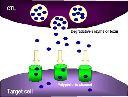
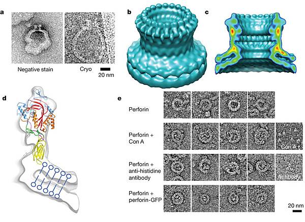
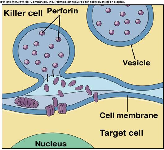
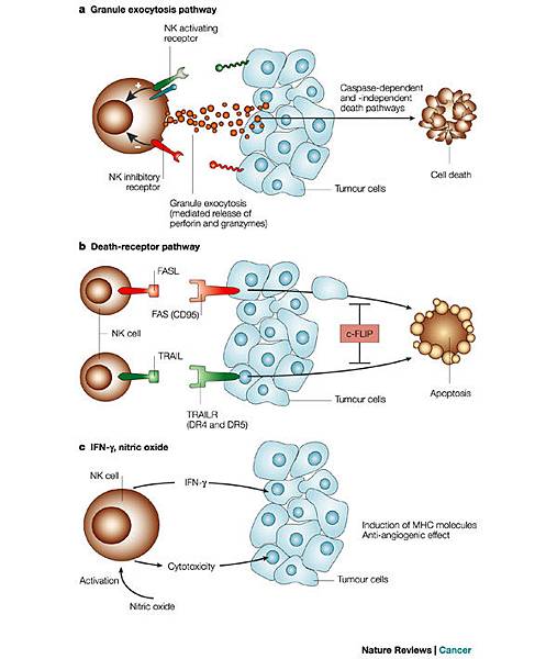
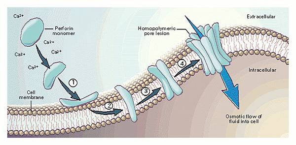
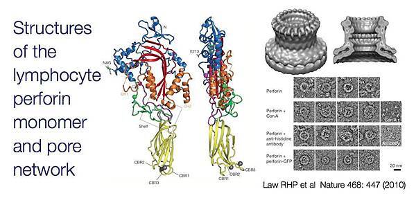
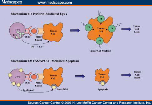
Perforin is a cytolytic protein found in the granules of Cytotoxic T lymphocytesCTLsand NK cells.

Charlie 發表在 痞客邦 留言(0) 人氣()