目前分類:Thread Lift 微整塑線拉皮 (164)

瀏覽方式: 標題列表 簡短摘要

281501615_m

20121229015217892s1_5386b03a19891  

25歲膠原蛋白流失高峰輕熟齡垂垮警訊

作者:健康醫療網/記者郭庚儒報導 | 健康醫療網 – 20141222

(健康醫療網/記者郭庚儒報導)

1339225561

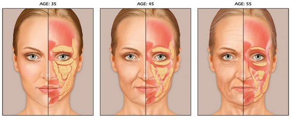
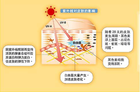
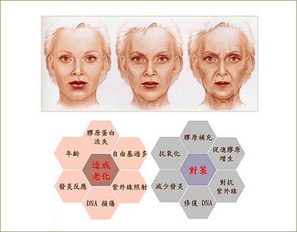
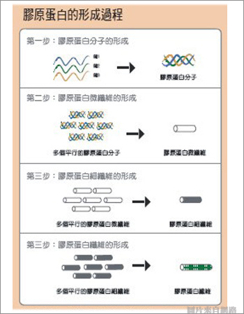
根據研究指出,肌膚於20歲起逐漸流失膠原蛋白,25歲後流失速度加遽當膠原蛋白流失速度大於生成速度,皮膚會失去彈性、出現各種皺紋,進而發生鬆弛下垂等現象。慶幸的是,目前醫學美容已發展出多種療程,如注射聚左旋乳酸、埋線或非侵入式的電波、音波拉皮等,能達到緊緻肌膚的目的,滿足愛美人士的需求。d344941

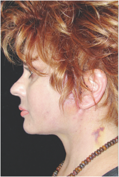
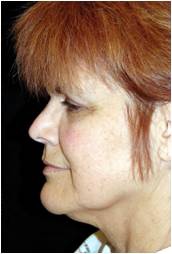
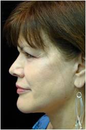
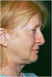
桃園一名35歲輕熟女,老化造成臉部鬆弛,有嘴邊肉、下臉脂肪堆積等問題,雖然曾做過電波拉皮但效果不明顯;日前,她經由朋友介紹得知玫瑰線拉皮療程,帶著試試看的心態,沒想到術後效果非常明顯,嘴角紋幾乎看不到了,皮膚也變得有彈性,讓她相當滿意。98

收治此個案的開業診所沈若蘭醫師指出,該名女子身材中等,但臉型圓短、下巴後縮,加上老化造成脂肪鬆弛下垂,下臉完全沒有輪廓線,看起來比實際體重胖幾公斤,經詳細諮詢與充分溝通後,在臉部兩側共埋了10條玫瑰線,術後不但改善下垂問題,整個人顯得年輕了,更有瘦了好幾公斤的感覺。

images

什麼是玫瑰線拉皮呢?沈若蘭醫師表示,玫瑰線是一種螺旋倒鉤的可吸收縫線,長約15~20公分,先在患者臉部的兩側耳際線或是髮際線開孔,各約0.1公分,將導管放入直達中下臉邊緣,再放入玫瑰線,每個孔可放34條,通常一邊68條,適合中下臉部鬆弛較嚴重、脂肪較多者,以及欲改善嘴角紋、法令紋、木偶紋者。

MegRyan-48-1998-2009

沈若蘭醫師指出,韓式埋線每條線約35公分,每埋一條線就要開一個針孔,13個月效果才會最明顯。而玫瑰線拉皮大多僅需2個孔,玫瑰線螺旋倒鉤的構造可直接勾住皮膚,透過醫師的手法往上拉提並固定,此外,線材也可刺激膠原蛋白生成修復,效果較快、傷口也較不明顯。

20100730091500616

若想要加強效果,沈若蘭醫師建議,可針對個人需求、鬆弛程度以及期望效果,玫瑰線拉皮可搭配不同的療程進行,包括注射玻尿酸或肉毒桿菌,以填補凹陷皺紋、改善動態性皺紋;或搭配4D埋線拉皮、電波拉皮、音波拉皮,都具有讓玫瑰線拉皮術後改善加乘的效果,尤其玫瑰線拉皮搭配音波拉皮,可深層收縮加上強力拉提。sub_c6_pic01

雖然玫瑰線拉皮效果較快速、滿意度高,不過,沈若蘭醫師仍提醒有意願進行療程的民眾,術前必須與醫師做好完整溝通,以達到雙方預期的術後效果,避免由於雙方認知的不同,而產生不如預期的落差。而糖尿病患者、有蟹足腫體質者、凝血功能不好者、嚴重過敏者、孕婦都不適合進行玫瑰線拉皮療程

1344673824-911957490

由於術後會有輕微的疼痛及腫脹感,可視情況冰敷或服用消炎止痛藥,傷口切忌碰水,約12天內可癒合。另外,1個月內禁止做臉、做誇張表情,清潔與保養臉部肌膚時,建議採用由下往上的手勢與手法,以免效果打折。

Charlie 發表在 痞客邦 留言(1) 人氣()

halslift

thread-lift-neck  

頸部回春方法多拉皮手術較有效

【記者蘇湘雲/台北報導】 | 台灣新生報 – 20141114


姿勢不當、頸部過度運動,小心頸部肌膚出現皺紋、下垂等問題!

楊志賢醫師表示,許多原因都會讓頸部產生皺紋、細紋。像頸部肌肉過度運動、長時間當低頭族,或者肥胖、頸部缺乏保養等因素都可能影響頸部外觀,因此平常就要多注意頸部保養。

楊志賢醫師指出,不少民眾只注重臉部保養,卻忽視頸部的保養。

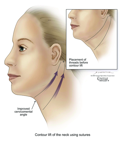
隨著年齡老化,頸部肌膚的真皮層結締組織會逐漸流失,進而開始產生鬆垮、下垂現象,如果頸部肌肉過度運動或長時間低頭滑手機,更容易造成靜態紋路,有時肥胖也會使肌膚加速產生皺摺,而上述因素都可能讓頸部肌膚看起來更加「滄桑」。


當頸部形成頸紋,就很難在短時間消失。

楊志賢醫師表示,民眾若想在短時間改善頸紋、頸部鬆垮、下垂困擾,就要考慮接受醫美治療。目前科技越來越進步,頸部回春方法也不斷推陳出新,像肉毒桿菌素、電波拉皮等都可以幫助改善頸部外觀。

肉毒桿菌素主要可以麻痺神經,讓肌肉不再持續動作、收縮,這樣就能暫時讓皺紋消失,而電波拉皮則是透過熱能促使膠原蛋白增生,進而讓肌膚更緊實。不過這些方法效果頂多維持半年到兩年時間,效果持續時間視每個人生活習慣、體質而有不同。

楊志賢醫師分析,當頸部出現淺層細紋,可以考慮以長效水凝膠填補下巴、木偶紋,如此便能改善淺層細紋。頸紋較嚴重時,就得考慮進行拉提手術,手術大多只需要局部麻醉,主要針對下巴、頸側、前頸等進行矯正、拉提。傷口切割點多隱藏於下頷上緣、下巴下緣,手術時間約一小時以內,這種治療方式改善頸紋效果較為持久。此外,接受手術後,建議可考慮運用「高壓氧」幫助新陳代謝、組織復原,並減少腫脹、疼痛。多數人大約六到八天即可進行術後拆線。


醫師提醒,術後暫時不要做彎腰、低頭、用力咳嗽等動作,才不會出血、腫脹。民眾想接受拉提手術一定要慎選醫師、醫療院所,減少感染風險。凝血功能異常、嚴重糖尿病患不宜接受手術。

Charlie 發表在 痞客邦 留言(0) 人氣()

tissue-adhesives-n-butyl-cyanoacrylate-tissue-glue-dermabond-500x500

F4.large  

整形膠立大功拉皮恢復期減半

作者:台北訊 | 中時電子報 – 2014714

中國時報【台北訊】

(趙欣報導)

對於歲月造成的肌膚嚴重鬆弛,已經無法透過微整形改善,只能依賴傳統拉皮手術才能真正找回年輕容貌,由於台灣的醫療品質與價格都具有優勢,目前不少選擇整形拉皮的病人都是專程從國外回來,利用短短停留台灣半個月左右的時間,就能帶著年輕十歲的樣貌,心滿意足的搭上回程飛機。


傳統拉皮手術需要三到四週才能消腫,這段期間因為臉部的血腫、瘀青,很多人根本不敢出門,而且消腫前還要在傷口擺放引流管,需很費心照顧。星光整型外科院長傅士榮表示,今年以來大部份的拉皮病人都會選擇搭配使用整形膠,大幅縮短術後照護時間,跟傳統拉皮手術相比,腫脹時間可以縮短一半以上,只需一週到十天左右就可以見人。


傅士榮醫師指出,對於一些台商或華僑來說,想要在有限時間內接受顏面整形手術,以改善歲月在臉上留下的刻痕,現在都會指定使用整形膠,一般來說只要在台灣待個十天左右,雖然臉上可能還有一點點腫,但是利用一點化妝品便可以修飾。

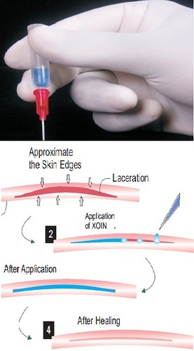
不像上臉拉皮可以從額頭以內視鏡進行手術,中下臉拉皮還是需要以傳統方式進行,傷口比較大,手術相對複雜許多,醫師需要打開整個皮瓣,難免有血腫的問題,但只要可以控制血腫狀況,瘀青跟疼痛問題就會大幅減輕。整形膠能快速將傷口組織凝血、密合,甚至能在幾分鐘內就形成生理性蛋白纖維架構,加速傷口的癒合過程,讓原本鬆弛的肌肉、筋膜與脂肪等軟組織,早日重新固定在拉皮後的位置上,臉部再度緊緻青春。


傅士榮醫師表示,隨著醫療技術進步,雖然拉皮手術的表皮傷口越來越小,但是皮下的傷口還是差不多大,目前中下臉拉皮的表面傷口會在耳前與髮際處,除非是年紀已經很大,需要進行頸部拉皮,才會需要從耳後進行手術。臨床觀察,進行拉皮手術的平均年齡在五十歲上下,其中男性佔了約一成,這些男性大部分都是因為還在職場衝刺,除了工作能力想要跟年輕人一較高下以外,外表方面也不想輸太多,因此在身體與經濟狀況都許可的情況下,就會來進行拉皮手術。

要注意的是,使用整形膠需要避免少數人對蛋白排斥過敏的現象,但是在國際文獻中甚少出現這樣的問題。拉皮手術與整形膠可說是目前整形最佳組合之一,只要注意手術後避免劇烈運動,前兩天最好冰敷,就能在最短時間內帶著青春新樣貌重出江湖。

Charlie 發表在 痞客邦 留言(0) 人氣()

QuickLift™Separating Fact From Fiction In Less Invasive Facelift Surgery3

COMPLICATIONSI often tell my patients that there is no real comfort in numbers because even if there is a 1or 2 percent chance of a certain complication, it is 100 percent if it happens to you. The reality of that is often difficult for the patient to understand. It is human nature to want to blame something or someone for a problem. However, with the complexity of the human body and the healing variables there within, complications WILL occur. It is not a subject that is typically dwelled on by patient or the doctor, since the chances of a significant complication are rare, but it is part of the patient’s decision and should be discussed before surgery.


Figure. QuickLiftTM With Significant Post-Procedure Bruising and Swelling; After Healing.

quicklift-preop2Discoloration days post-procedure.

quickliftweekslaterPatient same day with hair camouflaging area.

samedaypostQuickLift™Weeks later, after healing resolved.


In my experience, and in the medical literature, the most common complications occur within the first 48 hourshematoma, poor circulation (usually along the incision, which can lead to some scabbing and a wider healing scar or necrosis, also known as skin death), swelling and bruising. These complications are certainly manageable, and part of the process. In these situations, it is vital that the patient not panic. When the patient is completely compliant — listening to and following the physician’s instructions for follow-up care — it makes all the difference in the world.


Why I wrote this article

I want to close this article by explaining some important points about the benefits of less invasive procedures. Because we have developed a consistent, repeatable means of performing this surgical procedure, we are able to share it with other talented surgeons who can perform it in their own practices. In sharing the QuickLift™ with the profession, I am first training fellow surgeons in a specific medical procedure that, given the individual facts of a specific patient’s own medical condition, has proven over time to be repeatable from patient to patient, with great benefits to patients who are right for the procedure. But in three decades of practice, I’ve learned it’s not enough for doctors to talk to each other about procedures that can make a great difference in patients’ lives. You must also explain to patients why a particular procedure might be beneficial to them, and also, what risks or complications they might face. This is why I wrote this article. I want potential patients to understand the benefits and risks of this procedure, which could be beneficial to them. I want to help patients make the best choice for them. Aging is a fact of human existence. In the 21st century, in our society, good health, nutrition and modern medicine have us living longer, more fruitful lives. Yet far too many of us present a face to the world that doesn’t reflect how we feel on the inside, or the facts of our physical health. Less invasive facelift surgery such as the QuickLift™ can help us present a true picture of ourselves to the world.

If you are evaluating this kind of procedure for yourself, or for someone you love, I hope I have helped you to separate fact from fiction, and develop a true understanding of what you need to know about “quick” when it comes to facelift surgery.

facelift_visualcomparision1 

Figure. A Visual Comparison of Facelift Procedures.


Charlie 發表在 痞客邦 留言(0) 人氣()

QuickLift™Separating Fact From Fiction In Less Invasive Facelift Surgery2

We joke that gravity catches up with us. Like many jokes, there is truth in this one. As we age, we begin to sag at the jowls and around the eyes. The face we present to the world reflects less and less how we feel on the inside, the older we become.

This visible process of aging is more pronounced for women, who typically have thinner skin than men. It is not an old wife’s tale that women appear to age more quickly than men. It is scientific fact. In the first five years of menopause, for example, women’s skin accelerates from 1 percent annual loss in tonal quality to 6 percent a year, or 30 percent loss in those five years alone. After the first five years of menopause, the rate of decrease continues at 2.1 percent a year for women. While the changes for women may be more dramatic at first, the same aging process catches up with men, which is why 15-20 percent of all facelift patients today are men.

Thanks to modern cosmetic surgery, this rapid and sometimes startling change in the face we present to the world does not have to define who we are. We can make surgical and post-surgical changes in how we care for our face that can help us look as young as we feel on the inside. We can make changes in our appearance that spur us to make changes in our lives that lead to better health.

I have seen this time and again with my own patients — cosmetic surgery becomes the “signal point” or second chance in their lives to exercise regularly, lose weight, deal with stress, change jobs, and generally, take better care of themselves. Changing body image through cosmetic surgery can have profound impacts on how patients feel on the inside, improving mental and emotional health as well as physical appearance. Patients report that cosmetic surgery can make you feel better on the inside.


What’s the best type of surgery to present my “real” face to the world

The cosmetic surgery choice that is best for you will emerge only after you thoroughly investigate potential procedures, doctors, and outcomes. I advise my patients to make the decision that feels and looks right for them. I recommend that you listen to what others have to say. But listen most importantly to your own heart.

After nearly three decades of practice as a cosmetic surgeon, I speak from experience. That experience led me to develop the QuickLift™ and to continually work to perfect the procedure. I work with hundreds of patients each year who are good candidates for the surgery.

Early facelift procedures were limited in the extent of skin lifting to improve skin tone, and did not address the superficial musculoaponeurotic system (the SMAS, which is the covering of the facial muscles that I work with in performing the QuickLift). Consequently, the medical literature shows these early facelifts had a short life span, sometimes just a few years.


A Visual Comparison of Facelift Procedures

In the early 1990s, the composite rhytidectomy was introduced. This facelift procedure involves aggressive undermining of the face in a deep plane, and the elevations of the SMAS, cheek fat, and the muscles that close the eyelids (orbicularis oculi). Because of its more aggressive nature, this procedure typically has a higher complication rate, and patients can have a relatively long disfigurement period, with four to six weeks off of work and a period of six to 12 months before optimal results are achieved. What I have learned in three decades of practice is that patients with busy lives do not want to deal with prolonged swelling or a higher risk of complications. So when I think of “quick” with regard to the QuickLift™, certain aspects of the surgery come to mind. The most appealing aspect of this procedure is that it typically requires less downtime, meaning a patient can go back to their regular life more quickly. This begins with the surgery itself. It typically takes much less time to perform a QuickLift™ as compared to more extensive facelift surgery. Patients often experience procedure times of 90 minutes and sometimes less; more if adjunctive procedures are added. Second, because of the way the QuickLift™ is performed, patients typically need only local anesthesia, though additional sedative is sometimes indicated and offered by different surgeons. The potential risks, side effects, etc., all grow with more complex surgical procedures that require general anesthesia. And the medical literature shows that, the older we are, the more risk we face with general anesthesia. Third, the QuickLift™ procedure itself is less invasive medically. Traditional “deep plane” facelift surgery for instance, works at a deep skin level where your nerves lie. While this procedure may be best for some patients, the risk of nerve damage cannot be ignored. To work around the nerves, deep plane procedures require more surgical time, and more time in surgery typically calls for more intense anesthesia needs. The QuickLift, in contrast, is performed in the SMAS layer where there is little chance of nerve damage. In fact, of the thousands of QuickLift™ procedures performed nationally, I am unaware of any cases of permanent nerve damage. Put these important considerations together and it’s clear that if a particular kind of surgery takes less time to do, involves less aggressive anesthesia and less complicated surgical techniques, it is “quicker.” Less time in surgery, with a reduced need for anesthesia and less invasive techniques leads, generally speaking, too much quicker healing and results that are visible much sooner. This is an observable fact based upon thousands of successful procedures that have been documented in the medical literature.


No pain no gain

Even though the inference taken from Quick may be that the QuickLift™ is a drive-through procedure, it is vitally important to understand that the decision to have any surgical procedure, certainly a cosmetic one, is a big one. It is one that, regardless of the selected procedure and the quality of the care, may not lead to the expected outcome. In my title for this section, I did not mean to suggest that facial procedures are painful. By and large, they are not. However, the more aggressive the procedure, the more probable is the increase in the level of discomfort. Conversely, less aggressive typically translates into less discomfort. My point is that there are numerous issues that individuals go through in deciding to pursue facial enhancement.  Do they really want a facelift? What type of facelift is best for them? And what doctor do they choose to perform the surgery


Figure. QuickLiftTM With Minimal Swelling and Bruising Post-Operatively

quicklift-preopQuickLift™ Pre Procedure

quicklift-3days-postopQuickLift™ 3 Days Post Procedure

quicklift-7-days-post-opQuickLift™ 7 Days Post Procedure


Without going into great detail, here are some of the possible “curveballs” that may lead to a longer, or possibly, impeded journey to an expected outcome:

EXPECTATIONregardless of how well a doctor performs a facelift or QuickLift™, if the patient is expecting yin, and they get a yang, there will be some discontent. That is where consultation comes into play. Consultation is the ideal forum to create a realistic plan with the doctor in a private setting. What can reasonably be expected from the procedure relative to the facial conditions and goals of YOU, the patientNo procedure including the QuickLift™ is a cookie-cutter procedure. It must be customized to meet the various nuances that make each of our faces unique. This starts and often ends with the consultation.

Charlie 發表在 痞客邦 留言(0) 人氣()

QuickLift™Separating Fact From Fiction In Less Invasive Facelift Surgery1

January 12, 2010

What patients need to know about “quick” from the developer of the QuickLiftTM

By Dr. Dominic A. Brandy

https://quickliftmd.wordpress.com/category/quicklift/risks-of-the-quicklift/


Minimally invasive facelift surgery presents significant potential benefits to patients in terms of time spent in surgery, recovery periods and results. Understanding the benefits and risks of procedures such as the QuickLiftTM can help patients properly assess the suitability of this type of facelift surgery compared to other courses of action.

As the developer of the QuickLiftTM, a popular, less-invasive alternative to more complicated facelift surgery, I have worked with thousands of patients. I have also taught the QuickLift™ procedure to dozens of doctors. This has given me the opportunity to learn and understand the benefits and the risks of less invasive facelift surgery from the patient and the physician perspective. Having undergone the procedure myself, I have a personal connection to the patient as well as the physician perspective that I believe provides additional insight.

Seeing as many patients as I do, and hearing from as many doctors as I do, I feel that the true picture of facelift surgery is distorted in our digital age. It’s easy for individuals to misrepresent the true nature of facelift surgery in an exploding number of online forums, including blogs and video sites. It’s time for someone who knows the facts to put the record straight and provide the clear, honest information that potential patients seek.

I wanted to share the same information in this article that I share with patients in our practice. I want to separate fact from fiction and give a clear picture of the benefits and risks of facelift surgery. I know from personal experience (including my own procedure) that less invasive facelift surgery can produce good results. But I also know that some people have unrealistic expectations about cosmetic surgery. That is one reason I am writing this article, to put things in their proper perspective.


Does your face match your age

Let’s begin with an understanding of the need for facelift surgery. In the 21st century, the average health of a given man or woman is better than what it was 50 years ago. The good news is that as we continue to learn more about good health and nutrition, and combine that with modern medicine, we live longer. Internally, our organs, our bones, our muscles and our brains are generally healthier and last longer.

Until the advent of cosmetic surgery, we had not applied the same focus on the physical appearance of good health as we have to internal good health. Cosmetic surgery, including facelift surgery, allows us to look as good on the outside as we feel on the inside.

There is ample scientific support for cosmetic procedures that help retard the visible aging process. Our skin, generally speaking, is a fragile commodity. Scientists know that, from age 25 on, we lose about 1 percent of our skin’s tonal quality a year.

overtime-without-facelift 

Figure. The Faces of AgeAn Example of Aging Without Facial Rejuvenation

Charlie 發表在 痞客邦 留言(0) 人氣()

The Long-term Results of Purse-string Facelifts5

Conclusion

Over the years, I have tried to focus more on the perspective of my patients and less on my own. I now more clearly understand not only their physical appearance objectives, but also their concerns of risk, down time and cost; as well as their overall interest in an easier, less complicated experience. These desires have led me and other surgeons to develop procedures that are less invasive.

There has been skepticism expressed by some surgeons toward less invasive facelifts, which creates healthy dialogue, as long as it is based on fact and personal experience, not merely opinion. However, I believe the QuickLift and many purse-string mini-lifts are proving to deliver very good long-term results when the appropriate approach is used for the patient.

In general, my experience is that the S-lift works well on patients in their forties, that simplified MACS lifts and QuickLifts work well for patients in their fifties, that advanced QuickLifts (more anterior undermining and suspension) and MACS lifts (posterior cervicoplasty and malar suspension) work well for patients in their sixties, seventies and even eighties.

Charlie 發表在 痞客邦 留言(0) 人氣()

The Long-term Results of Purse-string Facelifts4

The QuickLift, like any other cosmetic procedure, is not a cookie-cutter solution. It must be applied differently to address each patient’s unique situation. Patient outcomes, as with every facelift procedure, will vary due to all of the physical characteristics mentioned, and those specific to the patient’s healing and intrinsic skin qualities.

Over the last six years, I have performed more than 1,800 QuickLift procedures. It has been my observation that the procedure produces consistent, long-term results (see figures 4a-b, 5a-b, 6a-b). I have also found that after the initial six to twelve month postoperative period, when virtually all of the expected skin relaxation has occurred, any further appearance changes are actually due to the normal aging process and not procedure shortcomings.


fig4aFig. 4a (before)  

fig4bFig. 4b (after)    

Fig. 4a-b: 64-year-old white female three years post-op from QuickLift, volume of brow and lips, and Hetter’s peel to the eyelids.


fig5aFig. 5a (before)  

fig5bFig. 5b (after)    

Fig. 5a-b: 67-year-old white female five years post-op from QuickLift and upper lid blepharoplasty.


figure-6aFig. 6a (before)  
figure-6bFig. 6b (after)    

Fig. 6a-b: 55-year-old white female five years and 10 months post-op from QuickLift and Hetter’s peel to the eyelids.

Charlie 發表在 痞客邦 留言(0) 人氣()

The Long-term Results of Purse-string Facelifts3

In the first 22 years of my career, I performed traditional superficial and deeper plane facelifts with results that I felt were good, but with complication and morbidity rates that were sometimes not acceptable.

In 1999, I heard Dr. Ziya Saylan present a lecture on the S-lift and started performing this technique on a select few patients with moderate, but encouraging results. I found that this technique worked well on patients in their forties, but for women who were post-menopausal (age 50 plus) the technique lacked the long-lasting improvement in the jowl and neck region that I and my patients sought. I felt that the 30 to 45 degree backward vector of the purse-string, insufficient undermining, and inferior platysmal tightening caused this result.


Developing The QuickLift

In 2003, I began changing the shape of the incision, increasing the degree of inferior-posterior suspension and the method of tightening (double concentric purse-strings) to create a more vertical 60 to75 degree advancement that I hoped would treat patients in their fifties, sixties and seventies more effectively.

In 2004, I published results from my first version of The QuickLift in the journal Cosmetic Dermatology. Since then I have modified the technique by increasing the length of the posterior incision and increasing the anterior undermining on certain patients who have heavier jowls, excessive submental redundancy and a sunken mid-face, which is often more present with older patients.

fig3 

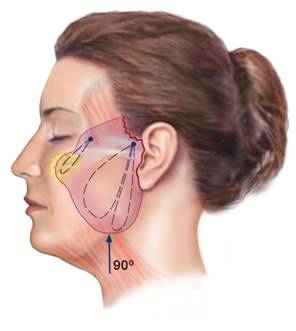
Fig. 3QuickLift. This schematic demonstrates double concentric overlapping purse-strings, incision (in red) and vector of advancements.


These changes create a more effective lift of the lower and mid-face regions, giving the surgeon more variety to treat these select patients more completely. Additionally, in a certain percentage of patients, I have modified the procedure with a posterior cervicoplasty and/or more anterior-inferior undermining.

Charlie 發表在 痞客邦 留言(0) 人氣()

The Long-term Results of Purse-string Facelifts2

The Procedures

Purse-string sutures are firmly anchored at either the zygomatic arch (S-lift and QuickLift) or at the temporalis fascia (MACS lift). The S-lift and the MACS lift use an O-shaped and a U-shaped purse-string. The MACS lift in some cases uses a third suspension purse-string to elevate the malar fat pad.

The QuickLift features a central, oval-shaped purse-string surrounded by an extremely large encircling purse-string. When purse-string suspension sutures are tightened, the SMAS is compressed, forming multiple gyri which then fibrose and give longevity to the procedure. In addition, as the purse-string is tightened, there is a pulley-type effect that strengthens the suspension with each additional throw of the suture and significantly reduces the chances of SMAS tear-through.

fig2 

Fig. 2MACS lift. This schematic demonstrates the undermining, O- and U-shaped purse-strings, malar fat pad purse-string, incision (in red) and vector of advancement.

Charlie 發表在 痞客邦 留言(0) 人氣()

The Long-term Results of Purse-string Facelifts1

February 26, 2010

https://quickliftmd.wordpress.com/category/quicklift/risks-of-the-quicklift/


Introduction

In recent years there has been a very strong trend toward less invasive facial rejuvenation. Patients want a cosmetic improvement but do not want to suffer prolonged recovery or a high risk of complication. Because of this, purse-string, suspension-based facelifts have gained great popularity.

However, many surgeons have voiced skepticism over the long-term effectiveness of these procedures. The three primary lifts that have been published in peer-reviewed journals are the S-lift (see figure 1), the MACS lift (see figure 2), and the QuickLift (see figure 3). All three techniques use the concept of suspending sagging facial features with strong, permanent purse-string sutures.

fig1 

Fig. 1S-lift. This schematic demonstrates the undermining, O- and U-shaped purse-strings, incision (in red) and vector of advancement.

Charlie 發表在 痞客邦 留言(0) 人氣()

Elevation of the Malar Fat Pad With a Percutaneous Technique6 

COMMENT

A simplified method of malar fat pad elevation is described. This method allows for elevation of the malar fat pad without extensive dissection. Complications were minor with this procedure when compared with endoscopic midface-lifting and complex deep-plane face-lifting procedures.

The procedure may be performed in conjunction with a face-lift (open technique), allowing for a more conservative face-lift technique than is customarily used to elevate the malar pad. It may also be performed without a face-lift to allow minimally invasive rejuvenation of the midface in the younger patient, in the older patient who has previously had a face-lift, or in the older patient whose aging is confined to the middle third of the face.


ARTICLE INFORMATION

Accepted for publication November 7, 2001.

This study was presented at the American Academy of Facial Plastic and Reconstructive Surgery Spring Meeting, Palm Desert, Calif, May 13, 2001.

Corresponding author and reprints: Gregory S. Keller, MD, 222 W Pueblo St, Santa Barbara, CA 93105 (e-mail: faclft@aol.com).


REFERENCES

1

Millard  DR  JrYuan  RTDevine  JW  Jr 

A challenge to the undefeated nasolabial folds Plast Reconstr Surg. 1987;8037- 46

2

Ivy  EJLorenc  ZPAston  SJ 

Is there a difference? a prospective study comparing lateral and standard SMAS face lifts with extended SMAS and composite rhytidectomies Plast Reconstr Surg. 1996;981135- 1143

3

Keller  GSCray  J 

Suprafibromuscular facelifting with periosteal suspension of the superficial musculoaponeurotic system and fat pad of Bichat rotation: tightening the net Arch Otolaryngol Head Neck Surg. 1996;122377- 384

4

Hamra  S 

The deep-plane rhytidectomy Plast Reconstr Surg. 1990;8653- 61

5

Psillakis  J 

Face lift without preauricular scars Plast Reconstr Surg.1994;941093- 1094

6

Owsley  JQ 

Lifting the malar fat pad for correction of prominent nasolabial folds Plast Reconstr Surg. 1993;91463- 476

7

Bosse  JPPapillon  J 

Surgical anatomy of the SMAS at the malar region Transactions of the Ninth International Congress of Plastic and Reconstructive Surgery. New York, NY McGraw-Hill Co1987;348- 349

8

Quatela  VCSabini  P 

Techniques in deep plane face lifting Facial Plast Surg Clin North Am. 2000;8193- 209

9

Anderson  RDLo  MW 

Endoscopic malar/midface suspension procedure Plast Reconstr Surg. 1998;1022196- 2208

10

Isse  NG 

Endoscopic facial rejuvenation Clin Plast Surg. 1997;24213- 231

11

Freeman  MS 

Endoscopic malar pad lift and subperiosteal midface lift Keller  GSedEndoscopic Facial Plastic Surgery. St Louis, Mo Mosby–Year Book Inc1997;109- 136

12

Hester  TR  JrCodner  MAMcCord  CDNahai  FGiannopoulos  

A Evolution of technique of the direct transblepharoplasty approach for the correction of lower lid and midfacial aging: maximizing results and minimizing complications in a 5-year experience Plast Reconstr Surg.2000;105393- 406discussion407- 408

13

Edelstein  CBalch  KShorr  NGoldberg  RA 

The transeyelid subperiosteal midface-lift in the unhappy postblepharoplasty patient Semin Ophthalmol.1998;13107- 114

14

Su  NN 

Closed suspension mini–cheek lift to reduce the nasolabial fold: a preliminary report Am J Cosmetic Surg. 1995;1231- 34

15

Sasaki  G 

Percutaneous suture elevation of malar fat pads  Paper presented at: Annual Meeting of the American Society for Plastic Surgery May 4, 2001 New York, NY Course 206

Charlie 發表在 痞客邦 留言(0) 人氣()

Elevation of the Malar Fat Pad With a Percutaneous Technique5 

RESULTS

At 3 months after surgery, all patients had significant elevations of their malar fat pads, with the exception of 2 early patients, who underwent revision surgery, with satisfactory results. The elevation was of an extremely natural appearance. Excellent volume replacement of the cheek region was obtained. Results, in the senior author's opinion, were improved over those seen in deep-plane face-lifting with malar fat pad fixation. The average malar pad elevation before creation of the temporal pocket was 2 to 3 mm. After the surgical procedure incorporated the creation of the temporal pocket, 4 to 5 mm of elevation was uniformly achieved.

Temporary asymmetries were seen in 8 patients, but resolved within the 3-month period. Temporary unilateral pain complaints were expressed by 3 patients, but also resolved within the 3-month period. Two patients, as mentioned above, underwent revision surgery to remove the palpable temple polytef bolster used in the early subcutaneous fixation technique.

All patients at 1 year after surgery (excluding the exceptions mentioned above) have maintained their malar elevation. These results are in contrast to those seen with deep plane face-lifting, after which the senior author has seen a regression of malar fat pad elevation in some of his (and other surgeons') patients. In fact, the technique provides an easy, minimally invasive "tune-up" in the malar area for rejuvenation of patients with previous face-lifts (Figure 5Figure 6, and Figure 7).

f 5 

Figure 5.

A, Preoperative photograph of 42-year-old patient with mild malar ptosis. B, Postoperative photograph taken 1 year after closed technique.

f 6 

Figure 6.

A, Preoperative photograph of 50-year-old patient with more-pronounced malar ptosis. B, Postoperative photograph taken 1 year after closed technique.

圖片7 

Figure 7.

A, Preoperative photograph of 49-year-old patient before surgery of the midface, jowls, and neck. B, Postoperative photograph taken after open technique (face-lift) and tip rhinoplasty. C, Preoperative oblique view. D, Postoperative oblique view.


No facial nerve problems, no excessive pulling, and no muscle fixation problems were noticed. There were spots of bruising in many patients, but they could generally be covered with makeup after about 72 hours. Four patients had prolonged infraorbital edema that required 2 weeks to resolve.

Charlie 發表在 痞客邦 留言(0) 人氣()

Elevation of the Malar Fat Pad With a Percutaneous Technique4 

The patient's hair is then braided out of the way of the incision, and the patient is prepared and fully draped. Meticulous attention to surgical technique is imperative to avoid infection. The surgeon uses a standard scrub and full gowning. Use of a modified sterile technique with surgical gloving and draping of the head area produced 2 surgical infections that required a course of antibiotics to resolve.

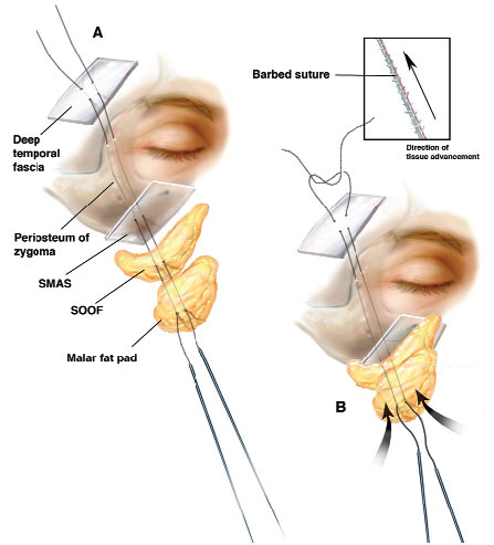
A No. 15 blade is used to make a 2- to 3-cm incision in the temporal hairline at the end of the projected pathway of needle travel. An iris scissors is used to complete the dissection down to the deep temporal fascia. An elevator is used to dissect a pocket posteriorly and then anteriorly past the brow along the deep temporal fascia in the pathway of needle travel. Creation of this pocket is a modification of the procedure that both protects the facial nerve and allows more elevation of the malar pad.

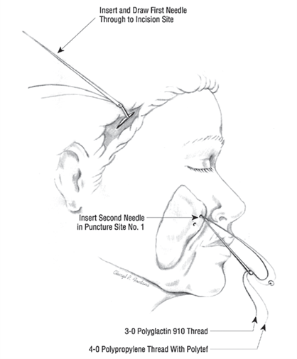
The surgeon then makes a small stab incision with a No. 11 blade at the demarcated spots that are to be the needle insertion points. The suture loop that was previously constructed with a Keith needle at each end is placed on the table. The Keith needle attached to each suture end is then passed through the stab incision to move the suture loop into position (Figure 3).

The Keith needle is inserted almost to the level of the bone and angled upward toward the temple incision. The plane of dissection is a palpable one that ends up in the "Suprafibromuscular" plane3 in the cheek, ie, the Superficial fascial plane of the face, Below the malar fat pad and Above the zygomatic muscles. An Aufricht nasal elevator is then placed in the temple incision, and the Keith needle is advanced into the temple area under the elevator and retrieved and pulled through the incision.

A "Sawing" motion with the 3-0 polyglactin 910 suture is used to create a "Passageway tract" for the 4-0 polypropylene–bolstered suture loop, which remains outside the stab incision. A release of the "puckered" tissue is then observed. This is the critical point of the operation, in that if the tract that is created is too shallow, a pucker can result. If the sawing motion is continued for too long and the tract is placed too deep, the subsequent elevation of the malar fat pad that is obtained will be too little.

If the tract does not result in a lack of a pucker or the desired elevation of the malar fat pad, the suture is then removed and another suture loop is placed.

The 3-0 polyglactin 910 suture is then removed. The 4-0 polypropylene–bolstered suture loop is then maneuvered through the stab incision and seated within the tract (Figure 4). Tugging on the suture loop demonstrates a lack of pucker and upward movement of the malar fat pad. The second suture loop is then placed through the second stab incision in a similar manner. After both suture loops are placed into position, a French eye needle is used to anchor the sutures to the deep temporal fascia. The sutures are then tied down and the temple incision is closed.

圖片4 

Figure 4.

Mobilization of the malar fat pad.

Charlie 發表在 痞客邦 留言(0) 人氣()

Elevation of the Malar Fat Pad With a Percutaneous Technique3 

While the surgeon is marking and injecting the patient, the assistant or scrub nurse is fashioning the double strand of sutures with a small polytef bolster. One end of a 4-0 polypropylene (Prolene) suture is passed through the eye of a Keith needle and stabilized with a hemostat. The suture is then passed through both ends of a 2 × 2-mm polytef bolster and passed through the eye of a second Keith needle and stabilized with a hemostat. Both ends of a 3-0 polyglactin 910 (Vicryl) or polyglycolic acid (Dexon) suture are then placed parallel to the 4-0 polypropylene sutures through the Keith needles and stabilized by the same hemostats (Figure 3).

圖片3 

Figure 3.

Needle passage.

Charlie 發表在 痞客邦 留言(0) 人氣()

Elevation of the Malar Fat Pad With a Percutaneous Technique2 

TECHNIQUE

The senior author has used 2 separate techniques. The first involved a superior subcutaneous anchoring of the suspension sutures in the temple via a subcutaneous polytefpatch (Gore-Tex; Gore-Tex Inc, Flagstaff, Ariz), as shown to him by Sasaki.15The sutures and polytef patch were anchored through a needle hole. No incisions in the temple or nasolabial fold were made. This technique was successful, but the senior author modified it because of several patients' ability to palpate the subcutaneous polytef patch. This annoyed 2 patients enough to require a minor reoperation, with removal of the patch and replacement of the sutures.

Our current malar pad suspension technique, which requires a small incision in the temporal hairline, involves fixation of the sutures to deep temporal fascia. With this technique, the patient is brought to the operating room and markings are placed as noted in Figure 2. These markings delineate the insertional points of the suture, the projected travel pathway of the Keith needle, and the temple incision and anchoring point.

圖片1 

Figure 2.

Markings.

Charlie 發表在 痞客邦 留言(0) 人氣()

Elevation of the Malar Fat Pad With a Percutaneous Technique1 

Gregory S. Keller, MD; Ali Namazie, MD; Keith Blackwell, MD; Jeffrey Rawnsley, MD; Sajjad Khan, MD

Arch Facial Plast Surg. 2002;4(1):20-25. doi:.

January 2002, Vol 4, No. 1 

Original Article | January 2002

http://archfaci.jamanetwork.com/article.aspx?articleid=479899


ABSTRACT

Objective  To describe a simplified method of malar fat pad elevation to rejuvenate the midface, nasolabial folds, labiomental folds, infraorbital hollows, and jowls via a percutaneous approach.

Patients and Design  One hundred eighteen patients were evaluated over a 12-month period in a prospective fashion by the operating surgeon. Preoperative and postoperative photographs were used for comparison.

Setting  Private practice.

Main Outcome Measures  Elevation of the malar fat pad by a measured amount.

Results  At 3 months, all patients had a significant elevation of the malar fat pad (3-7 mm), with the exception of 2 early patients who underwent revision surgery, with satisfactory results.

Conclusions  A simplified method of malar fat pad elevation is described. This method allows for elevation of the malar fat pad without extensive dissection. Excellent results are obtained with very little morbidity.


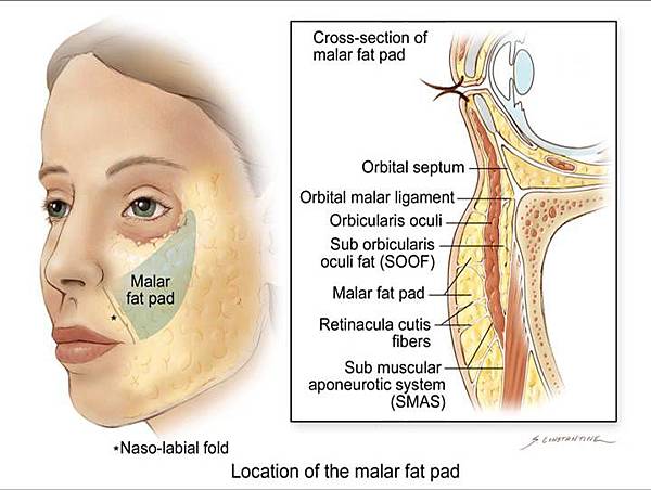
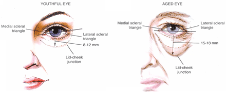
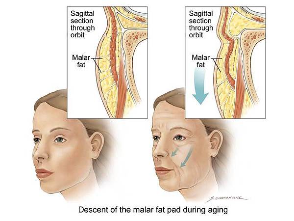
REVERSAL OF midfacial aging has become a focus of facial rejuvenation. Recently, the causes of midfacial aging have become well defined. The sagging of the Malar fat pad over the relatively fixed areas of the muscular and ligamentous connections of the nasolabial and labiomental folds produces a deepening of these structures. The downward migration of the fat pad produces hollowness in the midfacial and infraorbital areas that accentuates the aging process. Descent of the malar fat pad also contributes to the mid-face portion of the jowl (Figure 1).

 qoa10027f1

Figure 1.

The fallen malar fat pad produces midfacial ptosis, with an increase of the nasolabial and melolabial folds.


While midfacial aging is apparent in the older, classic face-lift patient, changes in the midface also appear in the 35- to 45-year-old patient who does not manifest other signs of aging. Many of these younger women, disturbed by the early signs of aging, seek classical face-lifts in an attempt to reverse aging confined to the midface.

Standard face-lifting techniques, associated with superficial musculoaponeurotic system plication, rotation, or resection, have resulted in only modest improvement of the nasolabial and mentolabial folds. The volumetric reduction of the fallen midface and infraorbital hollow associated with aging is also not fully responsive to these standard techniques.1-2 More aggressive techniques, such as deep-plane face-lifting, suprafibromuscular face-lifting, and subperiosteal face-lifting, appear to be more successful in addressing the midface in older patients, but are not widely adopted because of their increased morbidity and because they require advanced surgical training.3-8

In the younger patient, endoscopic techniques are used to avoid the surgical scarring associated with classic face-lifting. While successful, these techniques require complex instrumentation, sophisticated surgical technique, and a lengthy recovery period (that younger, working patients often do not have the time for). Most of them rely on suture elevation of the ptotic malar fat pad. Dissections and suture placement are difficult and/or associated with surgical morbidity.9-10 Surgeons also use techniques of malar fat pad elevation through a blepharoplasty incision. While useful for the correction of ectropian and eyelid contraction, these techniques have surgical morbidity and scarring in the lateral canthus.11-13

A simplified method of malar fat pad elevation to rejuvenate the midface, nasolabial folds, labiomental folds, infraorbital hollows, and jowls has obvious merits. Direct percutaneous suture suspension techniques to achieve these goals were attempted by the senior author (G.S.K.) and others over the last 10 years. While useful for stabilization of the malar fat pad, elevation of fallen structures was unable to be performed by direct suture without a pucker being produced at the site of suture placement.

In 1995, Su14 reported a successful technique for a "closed suspension mini–cheek lift to reduce the nasolabial fold," during which percutaneous suspension sutures are passed through the skin of the nasolabial fold with a Keith needle. The sutures are anchored to the deep temporalis fascia with a French eye needle after limited undermining with a closed-channel liposuction cannula.

Recently, Sasaki15 successfully elevated the midface with a direct percutaneous technique. Our 15-month results with a modification of this technique and sutures in 118 patients have confirmed its validity.

This percutaneous malar fat pad face-lift allows for reliable elevation of the midface with a relatively simple technique that can be performed by most skilled surgeons after they take part in an observational session, during which the critical parts of the procedure are elucidated. Surgical morbidity is minor. The procedure can be reversed, augmented, or modified in the postoperative period quite easily, in the event that changes are mandated or desired by the patient. It does not "change" a patient's appearance in the manner of a midfacial implant. It is also more reliable than autologous fat replacement and does not require multiple sessions to achieve a result.

The procedure is indicated in patients whose concerns of aging are confined primarily to the midface. Such patients are often younger, and are not ready for a face-lift. Males and older patients without excessive skin laxity (previous face-lifts, ethnic with elastic skin, etc) are also excellent candidates.

Charlie 發表在 痞客邦 留言(0) 人氣()

08

09

malarsep

tear_trough_blog3u78239674_130133ee5deg214charlize_theron1  

Anatomy of a "black eye"a newly described fascial system of the lower eyelid.

Clin Anat. 1998;11(3):157-61.

Pessa JE1, Zadoo VP, Adrian EK, Woodwards R, Garza JR.

http://www.ncbi.nlm.nih.gov/pubmed/9579587


Abstract

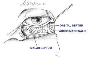
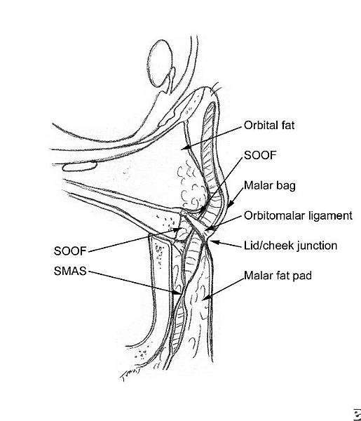
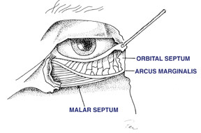
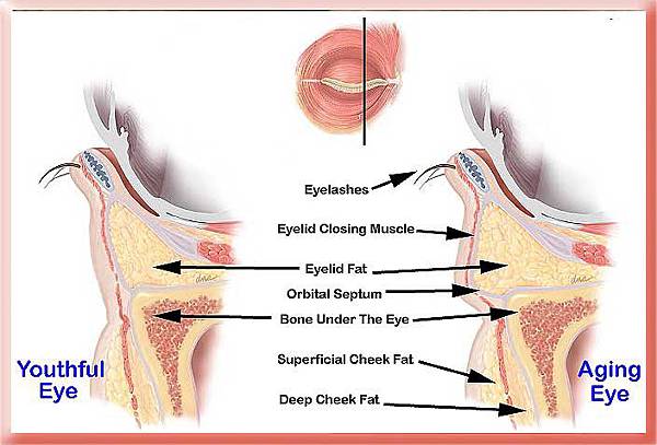
The anatomy of a black eye is examined in a series of cadaver dissections in which a previously unreported fascial system of the lower eyelid is identified. This fascia originates at the orbital rim, and is in continuity with the orbital septum and with the periosteum of the orbital floor and anterior maxillary wall. This fascia contributes to the thickened area along the orbital rim called the arcus marginale. At the level of the orbicularis oculi muscle, this fascia was noted microscopically to fuse with a fibrous septa of the superficial cheek fat. This creates one long continuous membrane from the orbital rim above to the cheek skin below. Dye injection techniques show that this membrane is impermeable and traps injected dye in the same place where a black eye forms. After periorbital injury, extravasated hemoglobin pigment is confined to the area above the cutaneous insertion of this membrane. This fascial system has been named the Septum malarismalar describes its origin along the orbital rim of the cheek, and septum further describes the partitioning nature of this ultra-thin membrane.

Charlie 發表在 痞客邦 留言(0) 人氣()

2008-03_02-02-large2008-03_02-01-large1378798889-27649410231378798898-3210262521Elevation-of-Malar-Fat-Pad

LO23_fig8LO23_fig6  

Midface lift of the malar fat padtechnical advances.

Plast Reconstr Surg. 2002 Aug;110(2):674-85; discussion 686-7.

Owsley JQ Jr, Zweifler M.

http://www.ncbi.nlm.nih.gov/pubmed/12142695


Most of the advances of the past decade in face lift technique have been directed to correcting the aging changes of the midface. With many midface lift techniques, patients typically experienced a prolonged period of periorbital ecchymosis and edema. Pessa's description of the anatomy of the malar septum has led to modifications of the senior author's (J.Q.O.) malar fat pad lift technique, designed to minimize postoperative ecchymosis and edema following mobilization of the malar fat pad. Preoperatively, markings of the cutaneous insertion of the malar septum are placed at the Infraorbital location. This line approximates the caudal margin of the musculus orbicularis oculi. Dissection under the malar fat pad is performed sharply under direct visualization. Care is taken to stay caudal to the malar septum insertion as marked on the skin. Refinements in the dissection of the midface malar fat pad have not altered the effective repositioning achieved. By leaving the malar septum intact, the amount of postoperative edema and ecchymosis is less than formerly associated with the dissection over the infraorbital orbicularis. With rare exception, patients were able to resume their usual activities and employment after a recovery period of 10 to 21 days postoperatively.

Charlie 發表在 痞客邦 留言(0) 人氣()

08091378798889-27649410231378798898-3210262521ghavami-lower_lid_five_step1malarseptear_trough_blog3  

Vertical Subperiosteal Mid-face-lift for Treatment of Malar Festoons6

Discussion

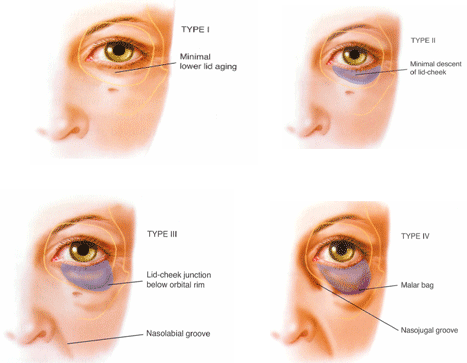
Festoons typically contain muscle and skin invaginations and must be differentiated from malar bags, which are edematous sack regions bordering on the cheek aesthetic unit that accumulate fat or fluid with age [1, 2]. Loose festoons of the orbicularis muscle are occasionally the cause of baggy eyelids, adding their bulk to that of sagging intraorbital fat [1, 2]. This is seen mostly in older patients who have lax supporting structures in the preseptal area, orbital area, and the jugal region of the lower lid [1]. The striated muscle fibers of the orbicularis muscle are constantly active and could exacerbate the attenuation of the orbicularis muscle in the aging process [1]. If the muscle contracts, the festoons are affected and reappear as the muscle relaxes. In rare cases a lateral-based transposition of the orbicularis muscle flap for orbicularis suspension in lower blepharoplasty is used for treating malar bags. With controlled amounts of traction to the lower lid, it is possible to correct the lowest part of the orbicularis oculi muscle due to its concentrating actions and its major vertical vector of traction. However, it is seldom effective in the treatment of festoons in the long run because it does not improve permeability characteristics of the malar septum.

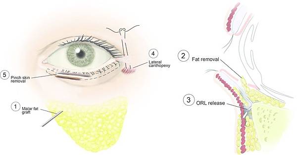
The question isWhich technique is most acceptable at the timeKnowing that gravity seems to have little effect on the development of malar mounds, the progressive distribution of skin and muscle due to chronic malar edema, malar festoon may be the end stage of the process [1, 2]. This is because the facial network originates at the level of the orbital rim and the malar septum acts as an impermeable membrane from the orbital rim to the cheek skin [2]. According to Pessa and Garza [2], the inferior border of the malar mound is created by dermal insertions of the inferior extent of the orbicularis muscle and that swelling in the malar mound region is primarily subdermal, forming festoons. Therefore, malar mounds tend to occur at a relatively constant location throughout life. Progressive distribution of skin results in the development of festoons because the facial fascia interdigitates with fibrous septa of the superior fascial cheek fat at the level of the orbicularis oculi as it crosses the muscles [2]. The malar septum, as described by Pessa and Garza [2], divides the Suborbicularis oculi fat into superior and inferior compartments. The inferior compartment is confluent with the lower cheek fat and the superior compartment contributes to the formation of malar mounds. The superior compartment contains the suborbicularis oculi fat, the orbicularis muscle, the superficial cheek fat, and the cheek skin. Thus, in some patients the malar mounds may be affected by fluctuating edema which can progress to festoons. Regarding the findings of Pessa and Garza [2], it seemed evident that Malar mounds, Edema, Festoons, and Periorbital ecchymosis all occur in the same anatomic area. That means that the cutaneous insertion point occurs roughly 3 cm inferior to the lateral canthus. Therefore, in agreement with Glasgold et al. [5], we do not believe that it is effective to use a subciliary incision alone to lift and redrape malar mounds or festoons; we believe that these are most effectively treated by direct excision of distributed skin [5]. Concerning our findings, malar festoons seem to be correctable only if the malar septum is affected. This can be achieved by a vertical subperiosteal midface elevation, release and reset of the malar septum, and a controlled amount of traction of the orbicularis oculi muscle in conjunction with little lower-lid skin resection. By vertically elevating the soft tissue of the cheek and thus repositioning of the malar septum, tissue edema (malar festoons) above its cutaneous insertion will be resolved.


Conclusion

Subperiosteal vertical midface lift resuspends and redrapes the facial network that originates at the level of the orbital rim and seems to improve permeability characteristics of the malar septum, which is the membrane from the orbital rim to the cheek skin. It resolves the malar edema, the malar mounds, and the loose festoons by freeing the cheek tissue from underlying bone and repositioning the malar septum.


Disclosures

J. F. Hoenig, D. Knutti, and A. de la Fuente have no conflicts of interest or financial ties to disclose.


Open Access

This article is distributed under the terms of the Creative Commons Attribution Noncommercial License which permits any noncommercial use, distribution, and reproduction in any medium, provided the original author(s) and source are credited.

Charlie 發表在 痞客邦 留言(0) 人氣()