目前分類:US-navigated MIS 超音波導航微創手術 (17)

瀏覽方式: 標題列表 簡短摘要

f_452637_1open versus laparoscopic surgery  

疝氣微創手術(Keyhole SurgeryMinimally Invasive Surgery)知多少?未知真相大揭露!

作者:健康醫療網/記者潘以慈報導 | 健康醫療網 – 20141114

(健康醫療網/記者潘以慈報導)

愛美是人的天性,所以在接受外科手術時,患者無不希望傷口越小越好,因此坊間有許多診所及醫院都標榜使用「微創手術」做為治療特色,強調術後僅會留下細小的傷口,其中包含疝氣治療;然而,卻有某些外科醫院在進行疝氣治療時假「微創手術」之名來吸引患者就診,卻行一般外科手術之實,只是將傷口由56公分縮小到34公分,而要患者自費。


醫界對於「微創手術」確實是有不同的認知。醫美手術方式慢慢朝著「疤痕越來越小、越來越不明顯」演進,之後這種手術痕跡較傳統手術較不明顯的手術方式,醫美界即泛稱為「微創手術」,這也是一般人對「微創手術」的認知。

根據維基百科定義,「微創手術是一種透過內視鏡及各種顯像技術,而使外科醫生在無需對患者造成巨大傷口的情況下施行的手術」。事實上,在醫療界中,真正關於腹腔內的微創手術(包括疝氣手術)指的是儀器進入腹腔的「腹腔內視鏡手術」。如果是進行疝氣手術的話,其方法是將病患全身麻醉後,灌注二氧化碳進腹腔使其漲大,再於腹部切三個11.5公分的小傷口使儀器進入腹腔,然後在疝氣部位使用人工網膜進行修補。


然而現在,坊間卻有某些醫院假「疝氣微創手術」之名,實際上卻行一般外科手術之實,並要患者自費的情形發生;事實上,這些醫院採用的並非真正的「腹腔內視鏡手術法」,而仍是以傳統的方式:在腹腔外切一傷口.以手術器材進入腹壁操作的手術。差別只是將原先需開56公分的傷口,勉強開小成34公分而已。疝氣專科醫師曾振橿表示,此舉會有一些不良影響,因傷口小,手術不易操作,容易拉長手術時間;助手需用力將傷口撐開,術後疼痛感劇烈;操作較難,對傷口內部組織傷害更大、腹壁也較不易確實修補導至復發率大幅提高。更重要的是,此方法與一般的傳統(非腹腔內視鏡)手術法並無差異,只因將傷口勉強開的較小,即自稱為「微創手術」,並比照「腹腔內視鏡手術法」收費。

因此,疝氣專科醫師曾振橿強調,民眾在接受疝氣手術前,應先確實瞭解各種手術法之差異,如果碰到手術方式及使用器材屬於健保給付範圍,但卻只因極小的手術結果差異(如單純手術傷口略小),就自稱使用新式或難度較高的手術方式,藉此巧立名目收取高額差價者,請謹慎評估其可信度,以免白花冤枉錢又沒得到真正安全的治療。


醫學小百科:

疝氣微創手術-「腹腔內視鏡手術」:病患須全身麻醉,且須導尿,並灌注二氧化碳進腹腔使其漲大,再於腹部切三個11.5公分的小傷口使儀器進入腹腔,然後在疝氣部位使用人工網膜進行修補術,可能需住院23天;此法因需全身麻醉,且手術時間長達12小時,是較危險的手術方式。

Charlie 發表在 痞客邦 留言(0) 人氣()

1E-HI8y-Omg85H4KX4xMDoxOjBrO-I4W8_10.620x360_q85_upscale

trigger_finger_nodule_gold_medF2.largegraphic-8.largekjae-56-60-g004-lmsk_image_2_-_500_x_trigger-finger-injectionTriggerFinger-Long

wp7deb9206_05_06wp8b87fb9b_05_06  

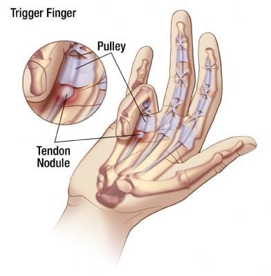
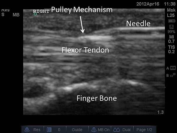
板機指(Trigger Finger)「喀」住!「手指屈肌腱的狹窄性肌腱鞘炎」多休息泡熱水緩解微創鬆解小傷口免縫合

作者:華人健康網記者張世傑/台北報導 | 華人健康網 – 2014621

常做家事,動作長時間反覆抓握重物,容易罹患板機指。


骨科門診中,「板機指」病灶很常見,醫師指出,這是一種手指彎曲如扣板機樣的疾病,主要是因為手指過度使用,造成肌腱發炎所致;千萬別以為板機指只有生產線員工才會有,事實上,包含電腦族、長期做家事等過度使用手指的動作都有可能罹患。


非關節病 主因肌腱鞘發炎

骨科蔡依樽醫師表示,「板機指」正式的學名是「手指屈肌腱的狹窄性肌腱鞘炎」,多數人常以為是指間關節出問題,其實是手指與手掌間的韌帶出問題,雖然五隻手指頭都有可能因為過度使用而罹患板機指,但最常見的還是大拇指、中指跟無名指

門診曾收治1名中年男性,由於工作需要長時間反覆抓握重物,導致手掌有一個局部突起疼痛點,手指一彎曲便會發出「喀」一聲,嚴重影響工作效率及日常生活,後檢查確診為板機指,經微創手術治療,讓手指恢復正常活動,不再手指卡卡。


注射類固醇 多休息緩惡化

事實上,板機指最直接的治療方法就是注射類固醇,通常超過一半的病患症狀可獲舒緩,因為類固醇有消炎止痛的效果,很快就覺得患部有改善,另外,也可同步進行熱敷、超音波等物理治療,但最好的方法還是減少手指過度使用,適度讓手指休息。


怕痛懶得動 常轉動易發炎

蔡依樽醫師強調,板機指好發於需要反覆彎曲手指、抓握動作的人,不少罹患「板機指」的民眾會因為害怕疼痛感,不然就是擔心彎曲動作會讓手指卡住,導致愈來愈不敢活動,如此長時間下來,反而會讓情況惡化。

另外,也有一種是相反的狀況,就是怕愈不動手指會不靈活,於是刻意去轉動手指或是不斷做伸展或彎曲的動作,這樣也會讓原有的患部形成發炎狀態。因此,最好還是請醫師判斷,並做最正確的診斷以及治療方法。


微創鬆解 傷口小免縫合

事實上,早期發現對症治療很重要,若置之不理或持續反覆發作,長期下來可能導致慢性發炎、通道過度窄縮,若治療效果不彰,就必須接受手術治。對於板機指的鬆解治療,目前最新的治療方式為微創鬆解法,僅需局部麻醉、傷口小(如捐血針孔、約0.1公分)、免縫合、免拆線,可縮短恢復期。


【醫師小叮嚀】:休息+泡熱水

板機指好發在5060歲的女性,而且女性的發生率是男性的26倍;預防及改善方法,就是要避免高頻率、長時間以及出力大的手指動作;如果已經有輕微症狀,除多休息外,可先做一些步驟改善不適,比方早晚將患部浸泡熱水,之後進行和緩的拉筋動作,這對於緩解症狀都有很好的幫助。

Charlie 發表在 痞客邦 留言(0) 人氣()

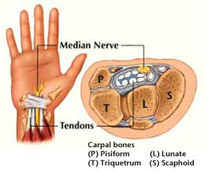
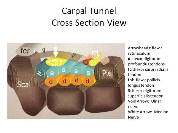
The safest location for steroid injection in the treatment of carpal tunnel syndrome.

Racasan O1, Dubert T.

J Hand Surg Br. 2005 Aug;30(4):412-4.


Abstract

Steroid injections are routinely performed for carpal tunnel syndrome. Direct needle injury of the median nerve is the major complication of these injections. The safest location of the injection remains controversial. The purpose of this study is to define safe guidelines to avoid nerve injury. The distances between the Median nerve, Palmaris Longus, Flexor Carpi Ulnaris and Flexor Carpi Radialis tendons were measured pre-operatively, 1cm proximal to the distal wrist crease in 93 endoscopic carpal tunnel releases. We found that the median nerve extended ulnarly beyond the Palmaris Longus tendon in 82 hands (88%). It is concluded that the median nerve is at risk if the injection is performed within 1cm on either the ulnar or radial side of the Palmaris Longus tendon. More ulnarly, there is risk to the ulnar pedicle. The safest location is to inject through the FCR tendon.

Charlie 發表在 痞客邦 留言(0) 人氣()

205264

carpal-tunnel-syndromeCarpal-Tunnel-Syndrome-picturewebmd_rf_photo_of_carpal_tunnel_illustration

WristCrossSectionShowingCarpalTunnel  

Comparison of needle position proximity to the median nerve in 2 carpal tunnel injection methodsa cadaveric study.

MacLennan A1, Schimizzi A, Meier KM, Barron OA, Catalano L, Glickel S.

J Hand Surg Am. 2009 May-Jun;34(5):875-9. doi: 10.1016/j.jhsa.2009.01.028.


Abstract

PURPOSE

Steroid injections are commonly performed by hand surgeons for relief of symptoms associated with carpal tunnel syndrome. The purpose of this study is to examine the relationship of the needle to the median nerve within the carpal tunnel and to the palmar cutaneous branch, using 2 injection techniques.


METHODS

Simulated carpal tunnel injections were performed on 15 cadaveric arms using 2 methods. The first injection used a widely accepted approach in which the needle is inserted at the wrist crease, just ulnar to the palmaris longus, and directed at a 30 degrees angle to the horizontal. In the second method, the needle is positioned just ulnar to the palpable ulnar border of flexor carpi radialis and angled 30 degrees to the horizontal. Specimens were dissected using an open carpal tunnel release. Calipers measured the distance from each needle to the median nerve within the carpal tunnel and to the palmar cutaneous branch.


RESULTS

Using the first injection method, the needle pierced the median nerve in 4 specimens, and its mean distance from the nerve measured 1.34 mm +/- 1.83 mm. With the second injection method, the median nerve was pierced in 1 specimen, and the needle averaged a distance of 4.79 mm +/- 3.96 mm from the nerve. In the first approach, the needle averaged 9.47 mm +/- 4.11 mm from the palmar cutaneous branch, compared to 1.74 mm +/- 1.59 mm with the second technique.


CONCLUSIONS

Physicians must exercise caution when performing carpal tunnel injections to avoid intraneural injection. The needle was a statistically significant shorter distance to the median nerve with the traditional injection method; however, the alterative method risks injury to the palmar cutaneous branch of the median nerve.

Charlie 發表在 痞客邦 留言(0) 人氣()

273v08nSupl.2-90178529fig10b63035cbb388eb9c3202bbb7cd1a5ecarpal-tunnel-illustrationctsus2Diagnostic-Ultrasound-of-Carpal-Tunnel-emedicinegraphic-1.largehqdefault

injrisk2Manos-PFR-ultrasound-img02

Normal Sonographic Appearance of the Carpal Tunnel

Paper_16029_abstract_3491_0ultrasound-image-of-carpal-tunnel

Sonographically guided carpal tunnel injectionsthe Ulnar approach.

Smith J1, Wisniewski SJ, Finnoff JT, Payne JM.

J Ultrasound Med. 2008 Oct;27(10):1485-90.


Abstract

OBJECTIVE

The purpose of this report is to describe a new sonographically guided technique for carpal tunnel injections using an Ulnar approach.


METHODS

Previously published sonographically guided techniques for carpal tunnel injections were reviewed. Described approaches were noted to be technically challenging because of the need to perform long-axis imaging of the carpal tunnel, short-axis (out-of-plane) imaging of the needle, or both.


RESULTS

We developed and herein describe the ulnar approach for sonographically guided carpal tunnel injections. Advantages of this approach include transverse imaging of the carpal tunnel, long-axis (in-plane) imaging of the needle, and versatility in targeting structures within the carpal tunnel.


CONCLUSIONS

Clinicians should consider the ulnar-sided approach when performing sonographically guided carpal tunnel injections.


Charlie 發表在 痞客邦 留言(0) 人氣()

2-Mapping-the-Hand-2

Incisionless-Carpal-Tunnel-Release-Surgery-Coda-CTRManos-AAOS-Hand-Trainer_0

MANOS_surgery_explained-300x226  

Ultra-minimally invasive sonographically guided carpal tunnel releaseAnatomic study of a new technique.

Rojo-Manaute JM1, Capa-Grasa A, Rodríguez-Maruri GE, Moran LM, Martínez MV, Martín JV.

J Ultrasound Med. 2013 Jan;32(1):131-42.


Abstract

OBJECTIVES

The purposes of this study were to measure a safe zone and a path for ultra-minimally invasive sonographically guided carpal tunnel release with a 1-mm incision in healthy volunteers and then test the procedure in cadavers.


METHODS

First, a previously reported Sonographic zone was defined as the space between the Median nerve and the closest Ulnar vascular structure. Axially, the safest theoretical cutting point for carpal tunnel release was set by bisecting this zone. Magnetic resonance imaging was used for axially determining the limits of the sectors (origin at the cutting point) that did not enclose structures at risk (arteries and nerves) and coronally for determining whether our release path could require directions that could potentially compromise safety (origin at the Pisiform's proximal pole). Second, in cadavers, we performed ultra-minimally invasive sonographically guided carpal tunnel release from an Intracarpal position through a 1-mm Antebrachial approach. Efficacy (deepest fibrous layer release rate), safety (absence of neurovascular or tendon injury), and damage to any anatomy superficial to transverse carpal ligament were assessed by dissection.


RESULTS

All 11 of our volunteers (22 wrists) had safe axial sectors located volar and radially of at least 80.4º (considered safe). Release path directions were theoretically safe (almost parallel to the longitudinal axis of the forearm). In 10 cadaver wrists, ultra-minimally invasive sonographically guided carpal tunnel release was effective (100% release rate) and safe without signs of intrusion into the superficial anatomy.


CONCLUSIONS

Ultra-minimally invasive sonographically guided carpal tunnel release in a safe sonographic zone may be feasible. The technique preserves the superficial anatomy and diminishes the damage of a surgical approach.

Charlie 發表在 痞客邦 留言(0) 人氣()

2-Mapping-the-Hand-2

Coda-CTR-procedure-ultrasound-structuresIncisionless-Carpal-Tunnel-Release-Surgery-Coda-CTR

MANOS_surgery_explained-300x226

Manos-AAOS-Hand-Trainer_0MANOS-CTR-Procedure-Surgeon-cutting-transverse-carpal-ligament-flexor-retinaculum2Manos-PFR-ultrasound-img02Manos-Superior-Distal-view-deployed-with-cap  

Ultrasound-guided carpal tunnel release using the manos CTR system.

Buncke G1, McCormack B, Bodor M.

Microsurgery. 2013 Jul;33(5):362-6. doi: 10.1002/micr.22092. Epub 2013 Feb 18.


Abstract

PURPOSE

Ultrasound (US) has been used in the management of carpal tunnel syndrome since the 1980s. The first report of US-guided carpal tunnel release (CTR) was published in 1997, with cadaver and clinical reports confirming the safe navigation of surgical tools with US for division of the transverse carpal ligament. The MANOS CTR device was recently reported as a minimally invasive tool for CTR, and may be well suited for use with US guidance.


PATIENTS AND METHODS

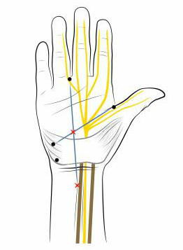
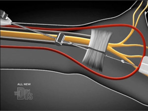
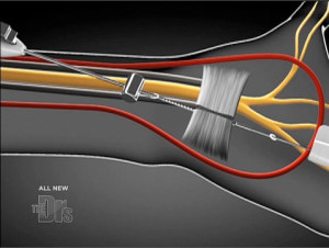
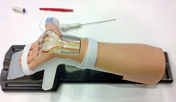
The authors report three cases of US-guided CTR using the MANOS CTR device. The MANOS device was inserted in a blunt configuration into the safe zone, and the cutting surface was deployed with a thumb-activated trigger that simultaneously ejected a sharp through the palm. The transverse carpal ligament was divided safely and confirmed with US.


RESULTS

US allowed for clear identification of the median nerve, safe zones, transverse carpal ligament, and the MANOS CTR device in relation to all pertinent structures of the carpal tunnel. Complete division of the transverse carpal ligament was confirmed in all three cases. There were no median nerve, vessel, tendon injuries, or chronic regional pain syndrome in any of the three cases.


CONCLUSIONS

US-guided CTR with the MANOS CTR device appears to be a safe technique and successful in confirming complete release.

Charlie 發表在 痞客邦 留言(0) 人氣()

Coda-CTR-procedure-ultrasound-structuresIncisionless-Carpal-Tunnel-Release-Surgery-Coda-CTR

manos_pfr_illus02

MANOS_surgery_explained-300x226

Manos-AAOS-Hand-Trainer_0MANOS-booth-backwall_550

MANOS-CTR-Procedure-Surgeon-cutting-transverse-carpal-ligament-flexor-retinaculum

MANOS-CTR-Procedure-Surgeon-cutting-transverse-carpal-ligament-flexor-retinaculum2Manos-Superior-Distal-view-deployed-with-cap  

Carpal tunnel release using the MANOS CTR systempreliminary results in 52 patients.

McCormack B1, Bowen W, Gunther S, Linthicum J, Kaplan M, Eyster E.

J Hand Surg Am. 2012 Apr;37(4):689-94. doi: 10.1016/j.jhsa.2011.12.033. Epub 2012 Feb 24.


Abstract

PURPOSE

To describe a carpal tunnel release technique using the MANOS Carpal Tunnel Release device, with preliminary results in 52 patients.


METHODS

The MANOS Carpal Tunnel Release device is a blade that divides the transverse carpal ligament using wrist and palm skin punctures. The awake patient provides feedback as the surgeon navigates a 2.1-mm-diameter blunt probe across the undersurface of the ligament from a wrist incision with standard disposable nerve stimulator monitoring. The leading tip of the blunt probe is uninsulated and conducts 2 mA. The surgeon converts the blunt insulated probe into an uninsulated blade by advancing a 0.9-mm needle through the palm with a thumb-activated deployment feature. The surgeon saws the ligament through the 2 skin punctures. We used a validated outcome questionnaire to assess postoperative symptoms at 3 months.


RESULTS

Symptom severity and functional status scores compare favorably with literature controls for open and endoscopic surgery at 3 months. One patient required reoperation for incomplete release. There were no tendon or nerve injuries.


CONCLUSIONS

Preliminary results suggest the MANOS Carpal Tunnel Release device to be safe and effective.


TYPE OF STUDY/LEVEL OF EVIDENCETherapeutic IV.

Charlie 發表在 痞客邦 留言(0) 人氣()

617149.fig.002  

Optimization of Cannula Visibility during Ultrasound-Guided Subclavian Vein Catheterization via a Longitudinal Approach by Implementing Echogenic Technology

Stefanidis K, Fragou M, Pentilas N, Kouraklis G, Nanas S, Savel RH, Shiloh AL, Slama M, Karakitsos D - Crit Care Res Pract (2012)


Bottom Line

Access time (12.1s ± 6.5 versus 18.9s ± 10.9) and the perceived technical difficulty of the ultrasound method (4.5 ± 1.5 versus 7.5 ± 1.5) were both decreased in the EC group compared to the NEC group (P < 0.05).

ConclusionsEchogenic technology significantly improved cannula visibility and decreased access time and technical complexity optimizing thus real-time ultrasound-guided SCV cannulation via a longitudinal approach.


AffiliationRadiology Department, Evangelismos University Hospital, 10676 Athens, Greece.

http://www.hindawi.com/journals/ccrp/2012/617149/


ABSTRACT

Objective.

One limitation of ultrasound-guided vascular access is the technical challenge of visualizing the cannula during insertion into the vessel. We hypothesized that the use of an echogenic vascular cannula (EC) would improve visualization when compared with a nonechogenic vascular cannula (NEC) during real-time ultrasound-guided subclavian vein (SCV) cannulation in the ICU. Material and Methods. Eighty mechanically ventilated patients were prospectively enrolled in a randomized study that was conducted in a medical-surgical ICU. Forty patients underwent EC and 40 patients were randomized to NEC. The procedure was ultrasound-guided SCV cannulation via the infraclavicular approach on the longitudinal axis.

Results.

The EC group exhibited increased cannula visibility as compared to the NEC group (92%±3% versus 85 ± 7%, resp., P < 0.01). There was strong agreement between the procedure operators and independent observers (k = 0.9, 95% confidence intervals assessed by bootstrap analysis = 0.87 to 0.93; P < 0.01). Access time (12.1s ± 6.5 versus 18.9s ± 10.9) and the perceived technical difficulty of the ultrasound method (4.5 ± 1.5 versus 7.5 ± 1.5) were both decreased in the EC group compared to the NEC group (P < 0.05).

Conclusions.

Echogenic technology significantly improved cannula visibility and decreased access time and technical complexity optimizing thus real-time ultrasound-guided SCV cannulation via a longitudinal approach.


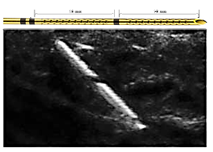
Fig2Echogenic cannula entering the SCV just adjacent to the sternum (A B); the former incorporates “Cornerstone” reflectors mainly arranged at its distal 2cm (C), which increase significantly its visibility (D).


MentionsAll patients were placed in Trendelenburg position and were cannulated as described in detail by Fragou et al. Triple-lumen catheters were used in all cases and all procedures were performed under controlled and nonemergent conditions in the ICU. Standard sterile precautions were utilized. The EC and NEC were both 18 gauge cannulas specifically intended for use in vascular access. Ultrasonography was performed with an HD11 XE ultrasound machine (Philips, Andover, MA, USA) equipped with a high-resolution 7.5–12MHz transducer, which was covered with sterile ultrasonic gel and wrapped in a sterile sheath (Microtec medical intraoperative probe cover, 12cm × 244cm). Using the infraclavicular approach, on the longitudinal axis, sonoanatomic landmarks (such as the acoustic shadows of the underlying first thoracic rib and of the sternum) were identified, as well as, the axillary and SCV vein (Figures 1 and 2). Doppler techniques were utilized to confirm the two-dimensional (2D) findings. Vessels were cannulated using the Seldinger technique under real-time ultrasound guidance.

Charlie 發表在 痞客邦 留言(0) 人氣()

Treatment of De Quervain's Syndrome with ultrasoundUS-guided infiltration of steroids and hyaluronic acid

http://posterng.netkey.at/esr/viewing/index.php?module=viewing_poster&task=&pi=122142

Congress : ECR 2014

Poster No. : C-2052

Type : Scientific Exhibit

Keywords : Outcomes, Inflammation, Puncture, Ultrasound, Percutaneous, Musculoskeletal system, Interventional non-vascular

Authors : L. Turturici1, E. Tarabelli2, R. Giuliani2, I. G. Burrelli2, P. Vagli1, P. Bemi1, C. Vignali2; 1Pisa/IT, 2Lido di Camaiore/IT

DOI : 10.1594/ecr2014/C-2052

DOI-Link : http://dx.doi.org/10.1594/ecr2014/C-2052


indexff1

Fig. 1 : The images show the location of pain and the course of the two tendons near the radial styloid process. References: U.O.C. Radiologia, Versilia Hospital, AUSL 12 of Viareggio, Lido di Camaiore/Italy 2013
indexff4

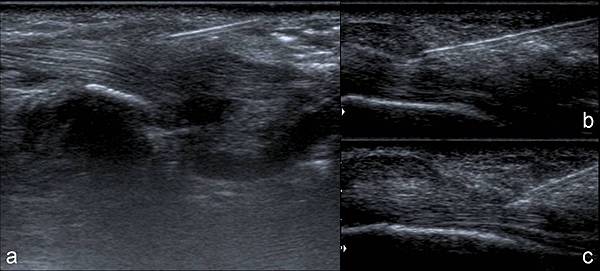
Fig. 4 : US monitoring during the procedure: immediately after the needle puncture local anhestesia is performed (a), the needle tip is inserted into the common tendon sheath (b), during steroids and hyaluronic acid injection a sheath distension (arrow heads) can be appreciated (c). References: U.O.C. Radiologia, Versilia Hospital, AUSL 12 of Viareggio, Lido di Camaiore/Italy 2013


Aims and objectives

To evaluate the effectiveness of US-guided infiltration of Hyaluronic acid (HA) and Steroids in the common tendon sheath of the Extensor pollicis brevis (EPB) and Abductor pollicis longus (APL) in the De Quervain's Syndrome in order to obtain a regression of the clinical status with functionality improvement and possibly avoiding a surgical intervention. De Quervain’s Syndrome is a Stenosing tenosynovitis of the First dorsal compartment of the wrist, affecting the EPB and APL tendons...


Methods and materials

34 patients with De Quervain's Syndrome were evaluated with US (fig.2) and treated with US-guided infiltration of HA and steroids. The procedure was performed percutaneously, using a sterile technique (fig.3), under US-guidance and with local anesthesia, puncturing selectively the common tendons sheath of the EPB and the APL with a 21G needle for the injection of steroids and low molecular weight (750 kDa) HA. US continous monitoring (with a 7-12 MHz linear transducer) depicted the...


Results

Three months after the procedure, a clinical improvement up to 80% reduction of QuickDASH score indexes was recorded in 28/34 patients (82.3%), while 6 patients (17.6%) had no regression of clinical symptoms and were retreated (tab.1). At 6 months follow-up 30 patients (88.2%) achieved a significant reduction of scores except for 4 patients (11.8%) that required a new treatment (tab.2). At 12 months follow-up an important clinical relief occurred in all patients, except 4 patients that...


Conclusion

Our data show that Selective infiltration under US-guidance of both Steroids and Hyaluronic acid in the common sheath of the EPB and APL provides a significative improvement of pain and function in the majority of patients affected by De Quervain’s Syndrome avoiding a possible surgery.


Personal information

Emilio Tarabelli MD, U.O.C. Radiologia, Versilia Hospital, AUSL 12 of Viareggio, Lido di Camaiore, Italy; emiliorx@libero.it  

Riccardo Giuliani MD, U.O.C. Radiologia, Versilia Hospital, AUSL 12 of Viareggio, Lido di Camaiore, Italy; giuliani@sirius.pisa.it  

Italo G Burrelli MD, U.O.C. Radiologia, Versilia Hospital, AUSL 12 of Viareggio, Lido di Camaiore, Italy; italoburrelli@yahoo.it  

Paola Vagli MD, Department of Diagnostic and Interventional Radiology.

Charlie 發表在 痞客邦 留言(0) 人氣()

Ultrasound-guided percutaneous treatment of rotator cuff calcific tendinitisrandomised comparison between one- and two-needle procedure

http://posterng.netkey.at/esr/viewing/index.php?module=viewing_poster&pi=121089

Congress : ECR 2014

Poster No. : B-0046

Type : Scientific Paper

Keywords : Musculoskeletal system, Interventional non-vascular, Ultrasound, Treatment effects, Calcifications / Calculi

Authors : G. Ferrero1, E. Fabbro1, D. Orlandi1, L. M. Sconfienza2, F. Lacelli3, G. Serafini3, E. Silvestri1; 1Genoa/IT, 2San Donato Milanese/IT, 3Pietra Ligure/IT

DOI : 10.1594/ecr2014/B-0046

DOI-Link : http://dx.doi.org/10.1594/ecr2014/B-0046


index1

Fig. 2: Calcifications: (a) Type I, (b) Type II, (c) Type III.

index2

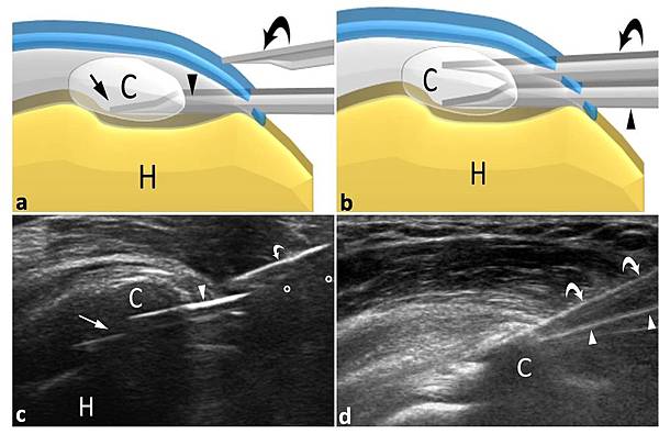
Fig. 11: A second needle (curved arrows) is inserted into the calcification (C) parallel...

index3

Fig. 13: A 20-ml syringe filled with warm sterile water is connected to one of the...


Purpose

CALCIFIC TENDINITIS OF THE ROTATOR CUFF Etiology

The term “calcific tendinitis” refers to the intratendinous deposition of Calcium, predominantly Hydroxyapatite, that can affect every tendon in the body and especially the rotator cuff. This pathological condition is a dynamic process that evolves through pre-calcific and calcific (formative, resorptive, reconstitutive) stages (Fig.1). In the precalcific stage, microtraumatic factors...


Methods and materials

IRB approval and patients’ informed consent were obtained. 100 patients (77 females and 23 males, mean age 46years, range 32-70y) with RCCT (single calcification, acute/post-acute phase, no tendon tears) were randomized into two groups and were treated with two different US-guided percutaneous tecniques. Group A (50 patients; mean visual analogue scale [VAS]=7.8) was treated using an US-guided 16G double-needle technique (local anesthesia, washing...


Results

The calcifications we treated in group A were: 4 fluid, 25 soft, 21 hard (mean dimension 23±5 mm). The calcifications we treated in group B were: 6 fluid, 27 soft, 17 hard (mean dimension 21±6 mm). We have had a drop-out of 3 patients from group B (2nd needle insertion). Overall procedure duration in group A (489±88s) was significantly shorter (P<.001) than in group B (684±187s). Difference in procedure duration in group A (489±88s ) was significantly lower than in...


Conclusion

Single- and Double-needle procedures are equally effective in treating RCCT with no major complications. Double-needle procedure allows for significantly reducing treatment time and appears to be much easier when dealing with soft and hard calcium deposits. Improvement in ease of calcium dissolution with double-needle technique when dealing with harder calcifications. Single needle could be dedicated to Acute phase and Double needle for harder Chronic phase.


Personal information

G. Ferrero, Department of Radiology, University of Genoa. (giulio.ferrero@gmail.com)

Charlie 發表在 痞客邦 留言(0) 人氣()

247_0

intralong_2

identifikation

intralong_3

MammaLock-SonoNeue_Sprotte_titelorganbiop

sprotte_kanuele

sprotte_spitze

Ultra-SonoPlex01Ultra-SonoPlex04Ultra-SonoPlex05

Ultra-SonoPlex02Ultra-SonoPlex03  

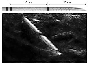
In-vitro visualization of biopsy needles with ultrasounda comparative study of standard and echogenic needles using an ultrasound phantom.

Hopkins RE1, Bradley M.

Clin Radiol. 2001 Jun;56(6):499-502.


Abstract

AIM:

To demonstrate the circumstances where echogenically enhanced biopsy needles confer an increase in visualization during ultrasound-guided biopsy using an in-vitro experiment.


MATERIALS AND METHODS:

An experiment using both standard and echogenically enhanced needles was conducted in a purpose built interventional phantom. The needle tip echogenicity and shaft visualization was recorded at different angles (20 and 60 degrees ) between the ultrasound probe and needle. The needle tip echogenicity was also recorded in four different angles of bevel rotation. The experiment was performed using a total of 18 different needles of 18 and 20 gauge.


RESULTS:

The experiment confirmed some previous findings. The needle was better visualised at an angle of approximately 60 degrees to the probe. The needle tip was optimally visualized with the bevel facing the transducer or 180 degrees to it. Needle tip movement improved conspicuity. Using an optimal angle of approach (60 degrees ) the use of an echogenic needle did not confer any increase in conspicuity. If the transducer to needle angle was suboptimal (20 degrees) then there was increased visualization of some echogenically enhanced needles.
 

CONCLUSION:

Optimisation of the conditions prior to ultrasound-guided needle biopsy will increase the visualization of the chosen device thus aiding the interventionalist. Needle gauge, bevel position, movement of the needle and the probe to needle angle all affect conspicuity. Needle coating or roughening does not improve tip visualization if the angle between the needle and probe can be optimized (60 degrees ). In unfavourable situations when the angle is suboptimal (20 degrees ), we have shown that echogenic enhancement of the needle tip can make it more visible.
Charlie 發表在 痞客邦 留言(0) 人氣()

b400727echoblock-echogenic-non-insulated-ultrasound-needles-6Echogenic-Tuohy-Needle-tip

echotuohy-echogenic-tuohy-epidural-needles-22

Einzeitige_techniken_header_SonoPlex_Stim_GBimage_largeNeedle-MicroLaser-Etchingsnewneedle1regular

SIG-Havel  

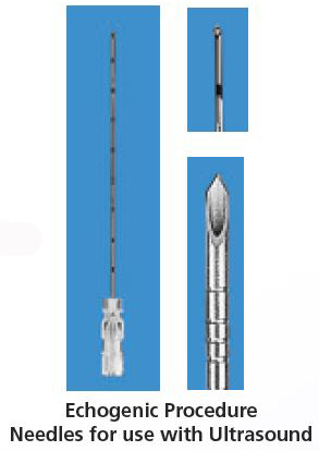
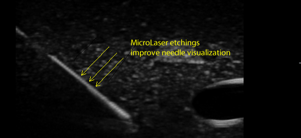
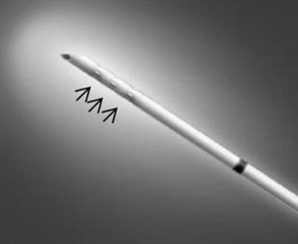
Needle echogenicity in sonographically guided regional anesthesiablinded comparison of 4 enhanced needles and validation of visual criteria for evaluation.

Sviggum HP1, Ahn K, Dilger JA, Smith HM.

J Ultrasound Med. 2013 Jan;32(1):143-8.


Abstract

OBJECTIVES:

Needle visualization is important for sonographically guided regional anesthesia procedures. Needle characteristics that improve needle visualization are therefore important to anesthesiologists. This study compared several echogenic needle designs by defining characteristics of needle echogenicity and assessing regional anesthesiologist preferences for these characteristics across various needle angles.


METHODS:

Twelve blinded regional anesthesiologists graded 5 randomized block needles (1 nonechogenic control and 4 echogenic) on 4 predefined characteristics (overall brightness of the needle, overall clarity of the needle, brightness of the needle tip, and clarity of the needle tip). In-plane needle images in a gel phantom were obtained at 4 needle angles (15°, 30°, 45°, and 60°). Participants rated specific needle characteristics for each needle at each angle and then ranked their overall needle preferences.


RESULTS:

Significant differences in all 4 needle characteristics were found across needle types (P< .01). Clarity of the needle tip was significantly associated with overall needle rank (P = .009). Other needle visualization characteristics were not significantly correlated with needle rank. The SonoPlex Stim needle (Pajunk Medical Systems, Tucker, GA) was rated highest in all 4 predefined needle characteristics as well as overall needle rank.


CONCLUSIONS:

This study shows that anesthesiologists prefer certain visual characteristics of needles used in sonographically guided regional anesthesia procedures. Specifically, needle tip clarity most closely predicted clinician needle preferences. These results support the idea that all echogenic needle designs do not uniformly enhance needle visualization. Further studies are needed to determine whether needles with superior tip clarity predict not only clinician preferences but also improved sonographically guided regional anesthetic outcomes.

Charlie 發表在 痞客邦 留言(0) 人氣()

235_Pajunk_704x212

AGHI_UNIPOLARI_NANOLINE_SONOPLEX_E_SONOPLEX_STIMEinzeitige_techniken_header_SonoPlex_Stimneedles

needles_1

Pajunk_Sonoplex_2up_a

clearid  

Echogenic technology can improve needle visibility during ultrasound-guided regional anesthesia.

Hebard S1, Hocking G.

Reg Anesth Pain Med. 2011 Mar-Apr;36(2):185-9.


Abstract

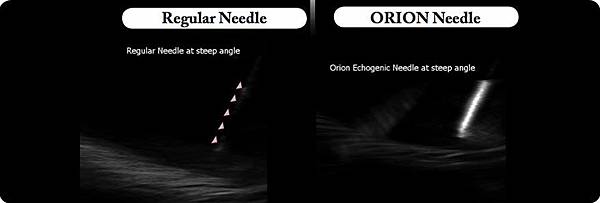
Needle tip visualization is fundamental to the safety and efficacy of ultrasound-guided regional anesthesia (UGRA). It can be extremely challenging especially at steep insertion angles. We assessed whether an echogenic needle improved tip visibility during UGRA by anesthesiologists performing their normal in-plane technique. The visibility of the Pajunk Sonoplex (echogenic) and the Pajunk Uniplex (control) needle were compared during 60 nerve blocks (30 femoral, 30 sciatic) in this randomized controlled trial. All ultrasound imaging was recorded for analysis. The anesthesiologist subjectively estimated the percentage time they had visualized the needle tip (5-point scale: 1 [0%-20%], 2 [20%-40%], 3 [40%-60%], 4 [60%-80%], 5 [80%-100%]). The actual time the tip was in view, angle of needle insertion, target depth, and procedure time were subsequently measured objectively by a single investigator. The Sonoplex group had both subjectively and objectively better tip visibility (P=0.002), despite having larger mean body mass index (29.0 vs 25.0 kg/m(2), P=0.01) and steeper mean insertion angle (31 vs 22 degrees, P=0.03). Objective percentage tip visibility, during in-plane UGRA, reduced by 12% for every 10-degree increase in insertion angle with the control. Tip visibility with the Sonoplex was independent of insertion angle over the range studied (0-57 degrees, P=0.95). This finding occurred when nonexpert anesthesiologists performed their standard UGRA technique. A needle that is visible for a greater percentage of time has potential safety and efficacy implications.


Copyright © 2011 by American Society of Regional Anesthesia and Pain Medicine

Charlie 發表在 痞客邦 留言(0) 人氣()

ULTRASONIC WELDING10-12-2011mobiusultrasoundvscanhcp201211006-02_300dpi1blanketgULTRASONIC WELDINGUltrasonicCut3  

Ultrasound and Ultrasonics

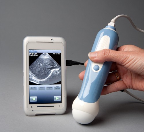
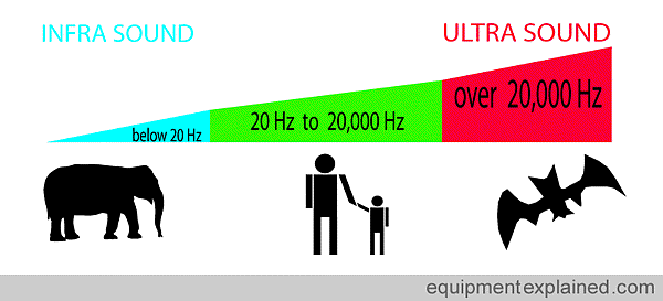
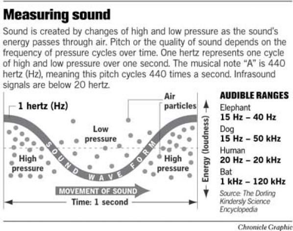
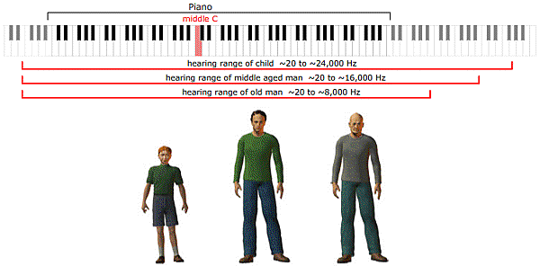
Ultrasound is an oscillating sound pressure wave with a frequency greater than the upper limit of the human hearing range.

Ultrasound is not separated from 'normal' (audible) sound based on differences in physical properties, only the fact that humans cannot hear it. Although this limit varies from person to person, it is approximately 20 kilohertz20,000 hertzin healthy, young adults.

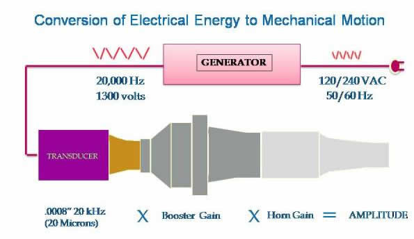
Ultrasound devices operate with frequencies from 20 kHz up to several gigahertz.

Ultrasound is used in many different fields.

Ultrasonic devices are used to detect objects and measure distances.

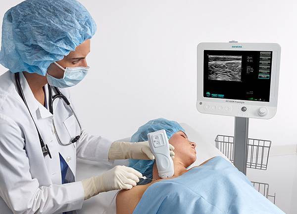
Ultrasonic imagingSonographyis used in both veterinary medicine and human medicine.

In the nondestructive testing of products and structures, ultrasound is used to detect invisible flaws.

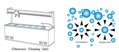
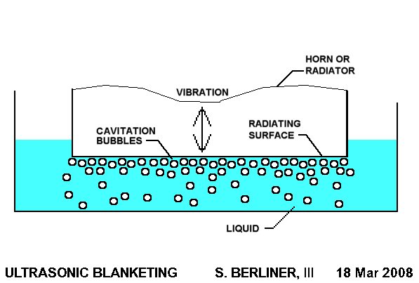
Industrially, ultrasound is used for cleaning and for mixing, and to accelerate chemical processes.

Organisms such as bats and porpoises use ultrasound for locating prey and obstacles.

Ultrasonics is the application of ultrasound. Ultrasound can be used for medical imaging, detection, measurement and cleaning. At higher power levels, ultrasonics is useful for changing the chemical properties of substances.

Charlie 發表在 痞客邦 留言(0) 人氣()

669267824_origquantifying_hearing_rangesnyu_soundwaves_activity1_figure1628x471source of infrasound

sources  

Infrasound and Infrasonics

Infrasound, sometimes referred to as Low-frequency sound.

Infrasound is Lower in frequency than 20 HzHertzor Cycles per second, the "normal" limit of human hearing. Hearing becomes gradually less sensitive as frequency decreases, so for humans to perceive infrasound, the sound pressure must be sufficiently high. The ear is the primary organ for sensing infrasound, but at higher intensities it is possible to feel infrasound vibrations in various parts of the body.

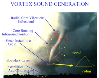
The study of such sound waves is referred to sometimes as Infrasonics, covering sounds beneath 20 Hz down to 0.001 Hz. This frequency range is utilized for monitoring earthquakes, charting rock and petroleum formations below the earth, and also in ballistocardiography and seismocardiography to study the mechanics of the heart.

Infrasound is characterized by an ability to cover long distances and get around obstacles with little dissipation.

Charlie 發表在 痞客邦 留言(0) 人氣()

Ultrasound-guided surgical drainage of face and neck abscesses

H. Yusa, H. Yoshida, E. Ueno, K. Onizawa, T. Yanagawa


Department of Oral and Maxillofacial Surgery, Institute of Clinical Medicine, University of Tsukuba, Tsukuba, Japan

Department of Metabolic and Endocrine Surgery, Institute of Clinical Medicine, University of Tsukuba, Tsukuba, Japan


International Journal of Oral and Maxillofacial Surgery

Volume 31, Issue 3, June 2002, Pages 327–329

Technical Notes


Abstract.

An ultrasound-guided surgical drainage technique in which grey-scale and colour Doppler ultrasonography were combined is described. The technique was performed for eight deep subcutaneous abscesses subsequent to odontogenic infection, and provided easy detection and accurate, reliable penetration of abscesses that were difficult to locate by physical examination. Colour Doppler ultrasonography is particularly useful for differentiating blood vessels from the static space of abscesses.


Keywords

ultrasound; grey-scale sonography; colour Doppler sonography; drainage; abscess

Charlie 發表在 痞客邦 留言(0) 人氣()