
Vertical Subperiosteal Mid-face-lift for Treatment of Malar Festoons(6)
Discussion
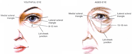
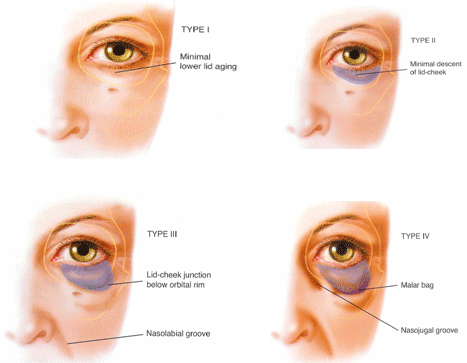
Festoons typically contain muscle and skin invaginations and must be differentiated from malar bags, which are edematous sack regions bordering on the cheek aesthetic unit that accumulate fat or fluid with age [1, 2]. Loose festoons of the orbicularis muscle are occasionally the cause of baggy eyelids, adding their bulk to that of sagging intraorbital fat [1, 2]. This is seen mostly in older patients who have lax supporting structures in the preseptal area, orbital area, and the jugal region of the lower lid [1]. The striated muscle fibers of the orbicularis muscle are constantly active and could exacerbate the attenuation of the orbicularis muscle in the aging process [1]. If the muscle contracts, the festoons are affected and reappear as the muscle relaxes. In rare cases a lateral-based transposition of the orbicularis muscle flap for orbicularis suspension in lower blepharoplasty is used for treating malar bags. With controlled amounts of traction to the lower lid, it is possible to correct the lowest part of the orbicularis oculi muscle due to its concentrating actions and its major vertical vector of traction. However, it is seldom effective in the treatment of festoons in the long run because it does not improve permeability characteristics of the malar septum.
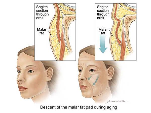
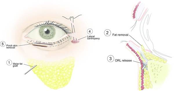
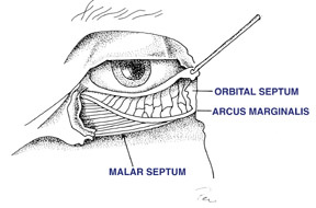
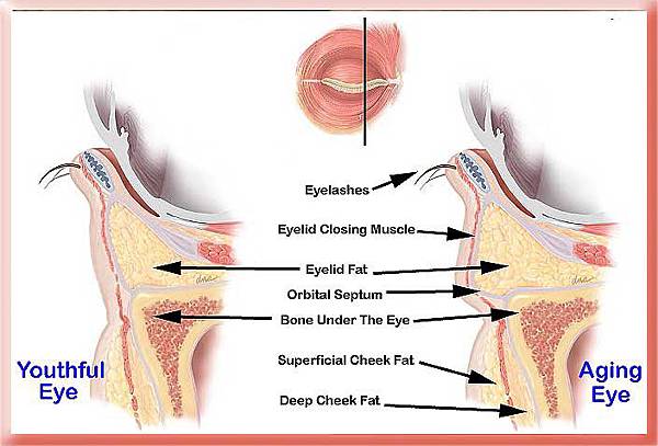
The question is:Which technique is most acceptable at the time?Knowing that gravity seems to have little effect on the development of malar mounds, the progressive distribution of skin and muscle due to chronic malar edema, malar festoon may be the end stage of the process [1, 2]. This is because the facial network originates at the level of the orbital rim and the malar septum acts as an impermeable membrane from the orbital rim to the cheek skin [2]. According to Pessa and Garza [2], the inferior border of the malar mound is created by dermal insertions of the inferior extent of the orbicularis muscle and that swelling in the malar mound region is primarily subdermal, forming festoons. Therefore, malar mounds tend to occur at a relatively constant location throughout life. Progressive distribution of skin results in the development of festoons because the facial fascia interdigitates with fibrous septa of the superior fascial cheek fat at the level of the orbicularis oculi as it crosses the muscles [2]. The malar septum, as described by Pessa and Garza [2], divides the Suborbicularis oculi fat into superior and inferior compartments. The inferior compartment is confluent with the lower cheek fat and the superior compartment contributes to the formation of malar mounds. The superior compartment contains the suborbicularis oculi fat, the orbicularis muscle, the superficial cheek fat, and the cheek skin. Thus, in some patients the malar mounds may be affected by fluctuating edema which can progress to festoons. Regarding the findings of Pessa and Garza [2], it seemed evident that Malar mounds, Edema, Festoons, and Periorbital ecchymosis all occur in the same anatomic area. That means that the cutaneous insertion point occurs roughly 3 cm inferior to the lateral canthus. Therefore, in agreement with Glasgold et al. [5], we do not believe that it is effective to use a subciliary incision alone to lift and redrape malar mounds or festoons; we believe that these are most effectively treated by direct excision of distributed skin [5]. Concerning our findings, malar festoons seem to be correctable only if the malar septum is affected. This can be achieved by a vertical subperiosteal midface elevation, release and reset of the malar septum, and a controlled amount of traction of the orbicularis oculi muscle in conjunction with little lower-lid skin resection. By vertically elevating the soft tissue of the cheek and thus repositioning of the malar septum, tissue edema (malar festoons) above its cutaneous insertion will be resolved.
Conclusion
Subperiosteal vertical midface lift resuspends and redrapes the facial network that originates at the level of the orbital rim and seems to improve permeability characteristics of the malar septum, which is the membrane from the orbital rim to the cheek skin. It resolves the malar edema, the malar mounds, and the loose festoons by freeing the cheek tissue from underlying bone and repositioning the malar septum.
Disclosures
J. F. Hoenig, D. Knutti, and A. de la Fuente have no conflicts of interest or financial ties to disclose.
Open Access
This article is distributed under the terms of the Creative Commons Attribution Noncommercial License which permits any noncommercial use, distribution, and reproduction in any medium, provided the original author(s) and source are credited.


 留言列表
留言列表
