1390989263139600577  

植髮技術大突破 美型植髮成為新趨勢

作者:健康醫療網/記者林怡亭報導 | 健康醫療網 – 2014129

(健康醫療網/記者林怡亭報導)

台灣植髮技術不斷突飛猛進,已超越國際水平,而且植髮技術已開始拋開過去傳統植髮技術的舊觀念,除了重視毛囊生存率以外,針對毛髮粗細度、毛髮密度、毛髮方向,都期許要與天生毛髮契合;在醫療與美學的結合下,進而慢慢衍生出美型植髮技術,竟然可以在眉毛、鬢角、鬍子、髮際線等部分移植毛囊,改變毛髮造型的需求,天然的植髮效果,已成為新一代植髮需求者所追求的新趨勢。

發毛診所林宜蓉醫師已經執行上千名美型植髮案例,她並於201310月,代表台灣到美國舊金山2013ISHRS國際植髮大會中,發表美型植髮變臉學術論文,引起世界植髪醫師關注,更受到很多國際知名植髮醫師的肯定,進而提升台灣植髮技術知名度,在全世界發光發熱。

林宜蓉醫師表示,美型植髮並非一蹴可及,美型植髮會按造患者的臉型,利用毛髮來改變造型,適用於眉毛、鬢角、鬍子、髮際線等部位,比起一般植髮技術還需要花更長的時間,植髮術前及植髮術中的設計,都需要花時間與患者做充分溝通後,才能進行美型植髮手術,植髮生存率應維持約8590%左右,考驗植髮醫師的經驗及技術和植髮團隊的協作,一氣呵成才能完成這巨大的美學工程。

台灣毛髮移植醫學會創辦人鄒積鎮醫師表示,民眾進行植髮手術前,應找合格醫師診斷,並對手術效果、風險、植髮資訊等充分溝通。務必要考量到診所的品質、醫師的專業技術及熟練度、手術的方法、毛髮生存及術後照顧等,避免因貪圖便宜植髮價格,而導致後悔莫及的窘境。再次提醒,民眾植髮可能一輩子只植一次,沒必要拿自己的頭開玩笑;醫療並非商業,最好選擇有口碑及ISO認證的植髮診所較有保障。

Charlie 發表在 痞客邦 留言(0) 人氣()

893080.fig.001893080.fig.002893080.fig.003best-lasers-to-treat-acne-scars

Acne scar classification

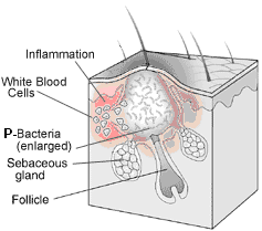
Acne scars are the result of inflammation within the dermis brought on by acne.

Acne scar is created by the wound trying to heal itself resulting in too much collagen in one spot.

Scarring can occur as a result of damage to the skin during the healing of active acne. There are two basic types of scar depending on whether there is a net loss or gain of collagen (Atrophic and Hypertrophic scars). Eighty to ninety percent of people with acne scars have scars associated with a loss of collagen (atrophic scars) compared to a minority who show hypertrophic scars and keloids.


Atrophic Scars

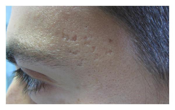
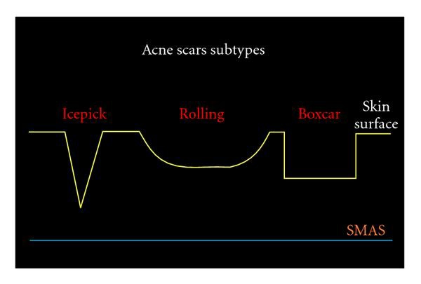
Icepick scarnarrow2mm, punctiform, and deep scars are known as icepick scars. With this type of scar, the opening is typically wider than the deeper infundibulum forming a ‘‘V’’ shape.

Rolling scardermal tethering of the dermis to the subcutis characterizes rolling scars, which are usually wider than 4 to 5mm. These scars give a rolling or undulating appearance to the skin‘‘M’’ shape.

Boxcar scarround or oval scars with well-established vertical edges are known as boxcar scars. These scars tend to be wider at the surface than an icepick scar and do not have the tapering V shape. Instead, they can be visualized as a ‘‘U’’ shape with a wide base. Boxcar scars can be shallow or deep.


The qualitative scarring grading system proposed by Goodman and Baron

Grades of Post Acne Scarring   Level of disease     Clinical features

  1. MacularThese scars can be erythematous, hyper- or hypopigmented flat marks. They do not represent a problem of contour like other scar grades but of color.

  2. MildMild atrophy or hypertrophy scars that may not be obvious at social distances of 50cm or greater and may be covered adequately by makeup or the normal shadow of shaved beard hair in men or normal body hair if extrafacial.

  3. ModerateModerate atrophic or hypertrophic scarring that is obvious at social distances of 50cm or greater and is not covered easily by makeup or the normal shadow of shaved beard hair in men or body hair if extrafacial, but is still able to be flattened by manual stretching of the skin if atrophic.

  4. SevereSevere atrophic or hypertrophic scarring that is evident at social distances greater than 50cm and is not covered easily by makeup or the normal shadow of shaved beard hair in men or body hair if extrafacial and is not able to be flattened by manual stretching of the skin.


Acne scar morphological classification

Acne Scars Subtype                       Clinical Features

Icepick scarIcepick scars are narrow2mm, deep, sharply marginated epithelial tracts that extend vertically to the deep dermis or subcutaneous tissue.

Rolling scarRolling scars occur from dermal tethering of otherwise relatively normal-appearing skin and are usually wider than 4 to 5mm. Abnormal fibrous anchoring of the dermis to the subcutis leads to superficial shadowing and a rolling or undulating appearance to the overlying skin.

Boxcar scar

Shallow

Diameter <3mm diameter

Diameter >3mm diameter     

Boxcar scars are round to oval depressions with sharply demarcated vertical edges, similar to varicella scars. They are clinically wider at the surface than icepick scars and do not taper to a point at the base.

Deep

Diameter <3mm diameter

Diameter >3mm diameter     

They may be shallow0.10.5mmor deep0.5mmand are most often 1.5 to 4.0mm in diameter.


Ice pick scarsDeep pits, that are the most common and a classic sign of acne scarring.

Box car scarsAngular scars that usually occur on the temple and cheeks, and can be either superficial or deep, these are similar to chickenpox scars.

Rolling scarsScars that give the skin a wave-like appearance.


Hypertrophic and Keloidal Scars

Hypertrophic scarsThickened, or keloid scars.


Charlie 發表在 痞客邦 留言(0) 人氣()

clip_image006wrinkle-maleskincare-age_aFitzpatrick 2FitzpatrickChartglogaumccallperez1  

Charlie 發表在 痞客邦 留言(0) 人氣()

Acne_t14

imgf000106_0001

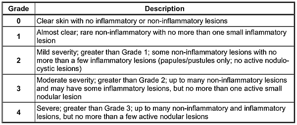
Acne Grading According to Burton Scale


Grade 0 - Total absence of lesions

Grade I - Sub-clinical Acne – few comedones visible only in close examination

Grade II - Comedonal Acne – Comedones with slight inflammation.

Grade III - Mild Acne – inflamed Papules with erythema.

Grade IV - Moderate Acne – many inflamed Papules and Pustules

Grade V - Severe Nodular Acne - inflamed papules and pustules with several deep nodular lesions.

Grade VI - Severe Cystic Acne – many modular cystic lesions with scarring.


Acne treatment should include a combination of the following resulting in the ability to maintain the achieved acne skin improvement over a twelve month period :

•an integrated methodology of patient education

•topical skin care

•topical acids


Charlie 發表在 痞客邦 留言(0) 人氣()

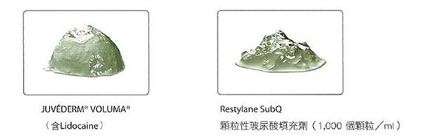
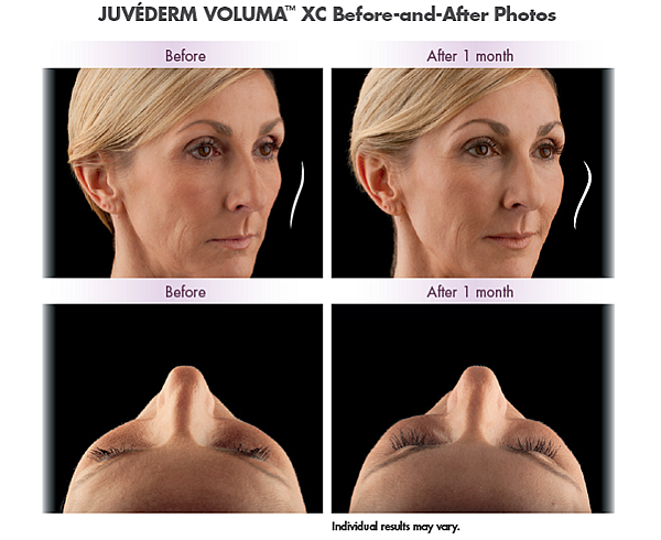
8-point-facelift604691_orig1380810707-3770630611380810733-4274098641380810747-33197009351380811011-25977735591380811024-4607099771380811056-15876788591380811456-32308302161380811468-22620279111380811478-39820320591380811487-40184478541380814254-24166001551381334424-2360601163Juvederm Voluma inj 20 mg_2 mL1200Picture_3-385x234Style-Your-Smile1VolumaBA1voluma-xcvolumaXCVolumaXClogo  

Charlie 發表在 痞客邦 留言(0) 人氣()

article-1

article-2article-2547481-1B0794E200000578-34_634x847

article-2547481-1B0794FB00000578-484_306x459

article-2547481-00461C4F00000258-953_634x484

The pretty TV presenter who deformed herself . . . for the sake of a “heart-shaped” face. Korean reporter undergoes brutal jaw surgery.

Before and after : The South Korean woman looks drastically different after undergoing plastic surgery

The young woman, allegedly a reporter on a TV channel, is one of thousands of Koreans who have had surgery.

Drastic moveThe woman is compared to a goblet in a photograph uploaded to a Japanese online forum.

The ideal: Many surgery-obsessed East Asian women strive to achieve the look of anime characters such as Sailor Moon, with a heart-shaped face and big eyes.

Southern Korean woman's drastic change shocks online forums.

She appears to have undergone jaw surgery to achieve 'heart shape' look

Unnamed woman follows continuously rising Korean surgery trend.


Charlie 發表在 痞客邦 留言(0) 人氣()

420_802e0ee8a7f63a09851f6ecfb98de0d8  

什麼叫「整壞了」 南韓電視台女記者舉例給你看

蘋果日報(施旖婕/綜合外電報導)

20140129  

韓國女記者整型前()和整型後()的差異。翻自英國《每日郵報》


南韓人整型成風,但南韓一名電視台女記者卻懷疑盲目整容,把原本姣好的面容給「整壞了」。

據英國《每日郵報》報導,該名女子「變臉前後照」最近被網友品頭論足。網友懷疑她在顎骨下功夫,試圖打造小臉,但效果弄巧成拙,下巴變得過尖,宛如外星人。有網友就認為,如此一來,整型反而把美女變成醜女,下巴更是一大敗筆。

Charlie 發表在 痞客邦 留言(0) 人氣()

x,qw=640,aq=80,av=1390551022200,asrc=http,P3A,P2F,P2Fs.nownews.com,P2F3c,P2Fd4,P2F3cd4c2a684b82c14e1997dfec78c21dc.jpg.pagespeed.ic.iJ6gt1Mm1Jxecb1503b28ab89a535d5381f19c4ce28.jpg.pagespeed.ic.oeYrwHd3xz

什麼叫「敗筆」?南韓女記者秀給你看 小V臉削削削!

Maggie Wu

NowNews 2014 01 29

國際中心/綜合報導

V臉、大眼娃就是南韓女性追求的變臉目標,不少人上整形診所削下顎,為的就是將國字臉削成小V臉,但這一位南韓女記者被網友批評好像削過頭了。(圖/翻攝自每日郵報)

網友諷過尖下巴像杯子。(圖/翻攝自每日郵報)


女性愛漂亮的程度恐怕是一般人無法想像的,而南韓人瘋整形的情況更是令人驚嘆,還記得先前的南韓小姐的選美大賽,各個佳麗卯起來大「鬥臉」,但卻被網友笑稱,根本分不出來誰是誰,就像複製人一般,可見南韓瘋整形的誇張程度。

根據英國《每日郵報》(Daily Mail)報導,小V臉、大眼娃就是南韓女性追求的變臉目標,不少人上整形診所削下顎,為的就是將國字臉削成小V臉,但這一位南韓女記者被網友批評好像削過頭了。

最近網路瘋傳一組「變臉前後照」,照片中的女子是南韓女記者,同樣也在顎骨下功夫追求終極小V,但是是否醫生下手過重,竟然一下子把下巴變得過尖,猶如外星人,引起網友抨擊。

有網友表示,整形原本是追求完美的臉型或身材,但有一些美女也盲目瘋整形讓自己變為醜女,甚至這位南韓女記者整過頭,讓下巴過尖簡直就是一大敗筆,反而沒有整還比較美!


Charlie 發表在 痞客邦 留言(0) 人氣()

Autotransfusion with RBC and Platelet-Rich PlasmaIntraoperative Autotransfusion2

3. QUALITY OF CELLS, ADVERSE EFFECTS

3.1. WASHED BLOOD

Cell separators remove Plasma from salvaged shed blood, eliminating potentially Toxic products of injured red cells and procoagulants <|[21]|>. Thus, the RBC concentrate is virtually Devoid of any particulate matter, including plasma proteins, coagulation factors and platelets (except for a small, clinically non-relevant fraction of nonfunctional cells and residual heparine) <|[10, 21]|>. Leucocytes are discharged to a varying degree, depending on the IAT device used <|[22]|>. Most of the Endogenous compounds released at the operative site and substances introduced by the surgeon are Washed out, as well as anticoagulants, antibiotics and other systemically administered drugs <|[23-25]|>. Conversely, some other endogenous Byproducts may be Concentrated (catecholamines, anaphylatoxines, leukotrienes, endotoxines) <|[25, 26]|>. Especially in orthopedic cases, wound blood frequently contains nonemulsified fat from bone marrow or subcutaneous depots which may ultimately cause venous fat embolism <|[11]|>. Devices based on discontinuous centrifugation cannot remove or filter out fat particles. Continuously processing cell separators are more effective in clearing fat and other contaminants such as inflammatory mediators <|[11, 14, 15]|>.

Excessive retransfusion of salvaged RBC and Saline may cause significant hematologic changes : Electrolyte and Acid-base balance may be disturbed by increase of sodium and chloride serum levels, and decrease of magnesium, ionized calcium, pH, pCO2, protein and albumine <|[25]|>. Extended IAT use may also cause a certain degree of hemolysis. While the consecutively augmented potassium release is counteracted by the washing process <|[11]|>, no cell separator can provide a complete clearence of Free hemoglobin, and serum levels may temporarily be elevated <|[8, 14, 15, 27]|>. Furthermore, since washed blood does not contain coagulation factors and platelets, massive RBC autotransfusion results in Dilutional coagulopathy and a Decrease of circulating platelets, requiring administration of the appropriate allogeneic components <|[6, 8, 19, 28]|>. Coagulation may further be disturbed by Prolonged hypothermia and shock, as well as by Reinfusion of residual heparin <|[1, 28]|>. Other adverse effects, such as Air embolism, Microembolism and Changes in 2,3-DPG affecting oxygen-carrying capacity of the red cells <|[8]|>, have become extremely rare if nonexistent after the arrival of electronically controlled, up-to-date cell separation devices.


3.2. UNWASHED BLOOD

Retransfusion of Unwashed autologous whole blood has been demonstrated to be safe and equieffective to Cell separation <|[16-19, 21]|>. However, the Quality of unprocessed blood is Inferior with a Higher return of lysed cellular debris, platelet microaggregates, fibrin split products, d-dimer, activated complement and free hemoglobin, indicating imbalanced coagulation due to pronounced hemostatic activation and increased hemolysis <|[Table I]|> <|[6, 19, 21, 27]|>. Especially in patients systemically anticoagulated for surgical purposes, simple IAT may not be detrimental in smaller amounts <|[6,19]|>, but larger quantities may lead to severe hemostatic disturbances with the potential of triggering Disseminated intravascular coagulation <|[6]|>. Because of this limited tolerance to unwashed wound blood, the total amount of retransfusion should clearly be restricted <|[6, 18, 21, 27]|>.


4. INDICATIONS

Intraoperative cell salvaging should be considered for all acute Emergency procedures. In severely traumatized patients with intense hemorrhage, the use of IAT may literally be life-saving due to a possibly limited supply of allogeneic blood to compensate for extreme blood loss. For practical purposes, the proper handling and processing of allogeneic blood transfusions in severe emergency situations may also pose substantial logistic challenges, thus supplemental autologous RBC supply provided by the comparatively simple IAT procedure may be extremely helpful. For these reasons, it is recommended that at least one IAT device should always be available and prepared in emergency rooms and operating units, and the attending staff must be well-trained in its proper use <|[4]|>.

In elective surgery, the routine application of IAT is controversial because of unpredictable cost-effectiveness <|[29, 30]|>. IAT has been shown to work efficiently in major orthopedic <|[9, 12, 31]|>, cardiac <|[32]|>, urologic <|[33, 34]|>, vascular <|[27]|>, intracranial <|[35]|> and gynecological <|[36]|> surgery and in emergency procedures, such as spinal trauma <|[37]|>. However, due to the wide variety of surgical, patient-related and institutional factors, it is difficult to define specific surgical procedures where IAT should always be used, and the overall blood salvage may not cover more than 25% of all perioperative blood losses <|[5, 12]|>.

<|[Table II]|> lists major surgical specialties where IAT can be successfully used and may be implemented in the perioperative blood saving strategies. As one practicable clinical guideline, the use of IAT is recommended for all major surgical procedures with anticipated blood loss of ≥ 1,000 ml <|[2, 5, 29]|>. At this level, IAT becomes cost-effective, but patients who are estimated to lose less than 1,000 ml are likely to receive little benefit yet incur substantial costs <|[29]|>. On the other hand, cost-effectiveness calculations limited to short-term perspectives do not take into account the enormous additional costs that may arise due to transmission of viral diseases or other adverse effects related to allogeneic transfusions <|[5, 38]|>.

Unfortunately, estimates of allogeneic blood requirement by preoperatively available data are very difficult and unreliable, even for planned, uncomplicated surgery <|[12, 39]|>. The anesthesiologists individual experience and knowledge of institutional circumstances (based on retrospective analyses), the type of procedure, and the patients red cell reserve, body mass and gender are yet the most helpful parameters in predicting intraoperative blood loss <|[12]|>. One rather elegant solution to this dilemma is the use of inexpensive intermediate storage bags whenever blood loss cannot be properly anticipated. Anticoagulated shed blood is put on hold, and when the amount of collected blood is sufficient to justify the use of IAT, it can be transferred to the cardiotomy reservoir and be further processed <|[4]|>. This strategy allows to save costs, while providing maximal patient safety.

Charlie 發表在 痞客邦 留言(0) 人氣()

550px-AUTOTRANSFUSIONPD

Fresenius CATS Diagram copy  

Autotransfusion with RBC and Platelet-Rich PlasmaIntraoperative Autotransfusion1

http://www.nataonline.com/node/433


Abbreviations

CPBcardiopulmonary bypass

PRPplatelets poor plasma

IATintraoperative autotransfusion

IPintraoperative plasmapheresis

RBCred blood cells


A. RBC

1. BACKGROUND SUMMARY

Intraoperative autotransfusion (IAT) is defined as the reinfusion of patient blood salvaged during and after surgery. IAT plays an important role in the context of perioperative blood saving strategies, having assumed standard of care status for many surgical procedures <|[1-6]|>.

The principle of IAT is to continuously collect intra- or post-operatively shed blood from the operative field. The salvaged blood is aspirated from the wound site and collected in a dedicated reservoir. Under standard conditions, Red cells are subsequently Separated, Washed, Hemoconcentrated and Stored for subsequent retransfusion to the patient. Only Erythrocytes are saved and retransfused, thus simultaneous volume and plasma replacement has to be provided, especially after processing of large quantities of shed blood <|[4]|>.

In contrast to stored RBC, freshly salvaged autologous red cells show Uncompromized functional capacity, oxygen delivery to tissues and survival, indicating that IAT has no significant detrimental effects on erythrocytes <|[7, 8]|>. IAT is most effective when combined with other autologous methods, particularly with Pre-operative autologous blood donation, Acute normovolemic hemodilution or Adiuvant drug therapies <|[4, 9, 10]|>.


2. METHODS

2.1. CELL SEPARATION (WASHED BLOOD)

This technique is based on Centrifugation, Separating red blood cells (RBC) from the Lighter components and fluids, including plasma, saline and buffy coat (Cell saver, Haemolite, Haemonetics, Braintree, MA; <|[figure 1]|>. Before starting the procedure, the system has to be filled with 100200 ml Heparinized saline (Priming), in order to Prevent cells from binding to membrane surfaces initiating Microaggregation, and to Diminish frictional forces and Damage to the cellular components <|[4, 8]|>. Blood released at the wound site is aspirated via a double-lumen suction catheter (80-100 mmHg), immediately anticoagulated (30,000 IU Heparin in 1,000 ml Saline) at the suction tip, and stored in a plastic cardiotomy reservoir, equipped with a microaggregate 120 m filter. When a minimum of 1,000 ml shed blood is collected, it is pumped into a rotating separation chamber (Latham bowl, 225 ml adaptable capacity), Washed with 10001500 ml Saline and concentrated. Whenever the extent or kind of surgical debris requires more extensive washing, processing cycles can be selected manually, and for emergency cases, washing can be skipped entirely. For pediatric patients, smaller centrifuges are available. As soon as the preset hematocrit is reached, the spinning separator chamber stops, and packed RBC, suspended in Saline solution, are pumped into an infusion bag, while the waste products are removed <|[4, 11]|>. After completion of each cycle, the bowl can be filled again for as many times as required. The Hematocrit of the final erythrocyte supension is regulated by an optical sensor in the centrifugation chamber, targeting 5570% of packed cell volume <|[4, 5, 11-13]|>.

Modified cell separation can also be performed by passing collected blood repeatedly through a vortex mixing filter with longitudinal channels and microporous plastic membranes (Haemocell System 350, Haemonetics), under continuous washing with saline solution <|[8]|>. Apparently, this less traumatizing method seems to have minor detrimental effects on the shed blood, better preserving the function of red cells and platelets <|[8]|>. However, no comparative studies have been performed up to now.

A recently developed novel device (Continuous AutoTransfusion System; Fresenius, Bad Homburg, Germany; <|[figure 2]|> allows continuous blood processing instead of operating in batches or units <|[11, 14]|>. The separation chamber used in this model represents a blood channel in the shape of a double spiral (capacity approx. 30 ml). Blood is pumped into the inner loop, while the separation chamber rotates continuously. Substances of less density leave the spiral immediately at this point, while RBC are moved towards the outer spiral, being continuously washed with saline <|[11, 14, 15]|>. All steps are performed simultaneously, allowing immediate retransfusion, even of smaller amounts, of processed red cells. Because of the maintained rotation of the separation unit, RBC cannot remix with lighter particles (eg. fat2). Thus, this novel cell separator may be more efficient in clearing substances that have been difficult to remove with conventional methods <|[11, 14]|>.

Disadvantages of cell separators might be less obvious : time and human resources are required for the setup, and the disposable single-use sets are expensive. For cases when blood loss may not be sufficient to warrant IAT operation, simple and inexpensive collection bags are available, allowing temporary storage of shed blood until further processing.


2.2. SIMPLE AUTOTRANSFUSION (UNWASHED BLOOD)

Retransfusion of Unwashed, Filtered whole blood (Srensen, Solcotrans, Biosurge) has been a clinically acclaimed alternative, both in regard to costs, ease of use and effectiveness in returning shed wound blood <|[16-19]|>. However, these devices have primarily been designed for the salvage of slowly oozing blood rather than rapid hemorrhage <|[8]|>. With the evolvement of new generations of safe, fully automated and easy-to-use cell separators, the importance of simple autotransfusion has largely shifted towards the postoperative phase <|[4, 6, 20]|>.

<|[Table I]|> summarizes typical properties of simple autotransfusion vs. cell separation devices <|[4]|>.

Charlie 發表在 痞客邦 留言(0) 人氣()