Vertical Subperiosteal Mid-face-lift for Treatment of Malar Festoons(3)
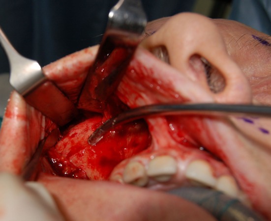
The soft tissue of the cheek is freed from underlying bone through a buccal incision placed from the molar to the canine to leave an adequate cuff of gingiva for closure (Fig. 4). Subperiosteal dissection is performed on the maxilla and zygoma using a custom-made periosteal rasp. The dissection extends from the nasal spine to the lateral buttress of the maxilla. It frees the tissue around the pyriform aperture, over the frontal process of the maxilla, along the inferior and lateral orbital rim, around the infraorbital nerve, then vertically up to the frontal process of the zygoma to the level of the lateral canthus, and laterally over the zygoma body to the zygomatic arch (Fig. 5). Freeing is performed over the anterior two thirds of the zygomatic arch. Release of the arcus marginalis is especially important for correcting the tear-trough deformity and elevation of the lid-cheek interface, just as freeing from the nasal spine and pyriform aperture is important for elevation of the upper lip and corners of the mouth.

Fig. 4
Intraoperative view of a patient undergoing a vertical upper-midface lift (SUM-lift); enoral view after degloving the maxilla, the zygoma, and two thirds of the zygoma arch. The suction cannula points to the sensitive zygomatic nerve

Fig. 5
Intraoperative view of a patient undergoing a vertical upper-midface lift (SUM-lift). The soft tissue of the cheek is freed from underlying bone through a buccal incision using a custom-made periosteal rasp. The dissection extends from the nasal spine ...
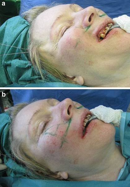
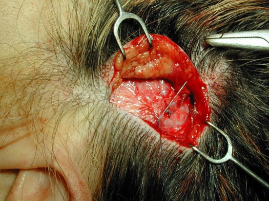
Once the soft tissue had been thoroughly freed, elevation of the midface begins by insertion of two 2 × 0 PDS sutures by an intraoral approach, taking a deep bite of the soft tissue (Fig. 6). The area for the cheek tissue suture fixation placement was marked previously; it is a cross in the medial cheek located at the intersection of a vertical line dropped from the lateral canthus and a transverse line directed from the lowest aspect of the alar groove at its intersection with the lip. The vector of suspension is determined entirely by the position of the key area relative to the point of bony zygomatic divergence. The patient’s head is turned to the side and a retractor is inserted through the short scalp incision into the temporal fossa. The patient’s head is straightened. The sutures are advanced up by suspending the cheek to the desired position until the identifying premolar show confirms adequate mobilization (Fig. 7a, b). The effects are assessed for symmetry and the sutures are fixed into the deep temporal fascia (Figs. 8 and and9).9). This superior vertical elevation lifts the deep tissues, which are maintained in the proper position of the fixed sutures. Most important is the production of a large soft tissue volumetric mass in the malar and submalar regions (Fig. 9). The distal end of the sutures with their multiple knots is trimmed and secured with a 4 × 0 Vicryl suture to lay flat in the temporal fossa, avoiding any discomfort of the sticky sutures.

Fig. 6
Intraoperative view of a patient undergoing a vertical upper-midface lift (SUM-lift). The area for the cheek tissue fixation placement is marked. It presents as a cross in the medial cheek located at the intersection of a vertical line dropped from the ...

Fig. 7a, 7b
Intraoperative oblique view of a patient undergoing a vertical upper-midface lift. The midfacial tissues are elevated until identifying premolars confirms adequate mobilization to the desired position by applying tension to the sutures exiting the ...

Fig. 8
Intraoperative view of a patient undergoing a vertical upper-midface lift. Superior vertical elevation produces a large soft tissue volumetric mass in the malar region. It is most important to assess large soft tissue volumetric mass production in the ...

Fig. 9
Intraoperative view of a patient undergoing a vertical upper-midface lift. After mobilizing the midface tissues and the SMAS fascia, the mesotemporalis fascia is repositioned, suspended after suture fixation, suspending the midface tissue to the deep ...
In addition, the frontotemporal skin is lifted in a vertical direction and is also anchored to the temporal fascia. Several interrupted sutures are secured to the temporal fascia at the level of the access incision, allowing an open knotting technique and secure fixation of the flap (Fig. 9). After performing the midface elevation, a laterally based transposition orbicularis muscle flap, described by Hamra [15, 16], is used (Figs. 10 and and11);11); it is a safe and effective method to transmit a controlled amount of traction to the lower lid in lower blepharoplasty without the need for canthoplasty or canthopexy. The loose, redundant lower eyelid is meticulously removed without any muscle fibers. After meticulous homeostasis, the buccal sulcus is closed with interrupted sutures.

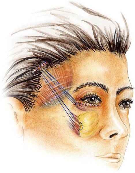
Fig. 10
Illustration of the vertical upper-midface lift with the suspension sutures in place. Note the suspension of the soft tissue at the inferior lateral orbital rim area.

Fig. 11
Intraoperative view of the suspension sutures placed in the soft tissue at the inferior lateral orbital rim area after dissection of the orbicularis muscle for septal release and muscle suspension.


 留言列表
留言列表
